
Presentation
Fall from bicycle resulting in an injury of the lower left hemithorax by the handlebar. The patient complains of a sharp pain in the chest, aggravated by deep breathing.
Patient Data

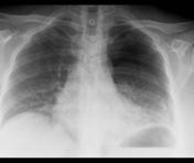
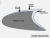
Note the visible visceral pleural edge and a relatively increased transparency of the involved left hemithorax on the supine AP radiograph. This occurs as in the supine position gravity causes the lung to fall dorsally allowing the air to collect anteromedially, laterally and basally.
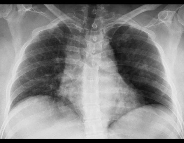
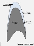
In the erect projection the large pneumothorax is far more evident since the lung is positioned more inferiorly. Thus, air is seen at the apex of the chest showing the familiar appearance of the visceral pleural line parallel to but separated from the chest wall.

Case Discussion
The case demonstrates traumatic pneumothorax caused by penetrating or blunt trauma to the chest.
The supine pneumothorax is best confirmed on full expiration, but imaging in an erect position is preferred if the condition of the patient allows it.
In this example the pneumothorax seems to be small in the supine projection, while the erect radiograph reveals its full extent (necessitating placement of a chest tube).


 Unable to process the form. Check for errors and try again.