
Presentation
The patient suffering from epigastric pain for a long time.
Patient Data
Age: 70 years
Gender: Female
From the case:
Peptic ulcer


Download
Info
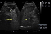
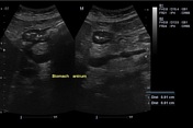
Stomach antrum wall thickness of 9.1 mm with a suspected lesion in the pylorus
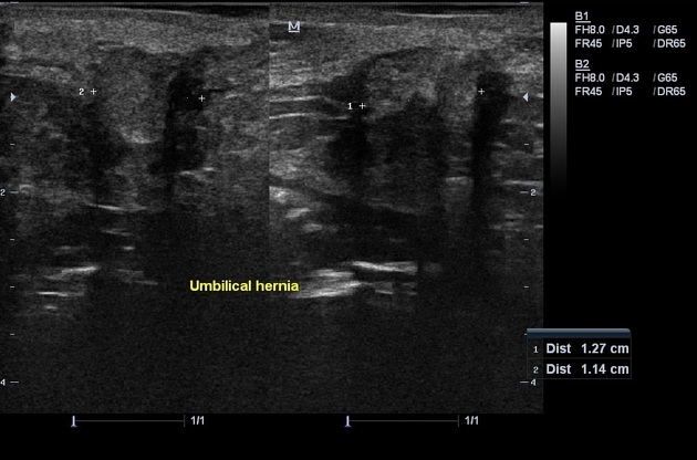
Umbilical hernia: 11 x 12 mm
Colon wall thickness: 7-8 mm
Minimal free fluid.
Endoscopy was done by Muneer ALrateb MD, Internist, Lattakia, Syria.
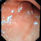
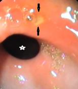
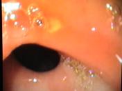
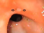
From the case:
Peptic ulcer
Download
Info

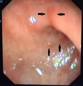
Acute antrum inflammation (arrows).
Peptic ulcer (yellow area surrounded by red contour) above the pylorus (star)
Case Discussion
Ultrasound of the stomach is not specific for stomach diseases, but maybe give us a flash.
It needs experience and clinical correlation, if any suspicious findings are noted, endoscopy and other investigations become recommended.


 Unable to process the form. Check for errors and try again.