Presentation
Acute diplopia.
Patient Data
Age: Adult
From the case:
Weber syndrome


Download
Info

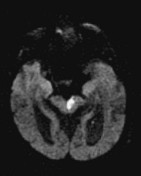
Selected images from an MRI demonstrate a left sided mid-brain region of high T2 signal with matching restricted diffusion. This is consistent with an acute stroke causing Weber syndrome.
Case Discussion
Weber syndrome is a midbrain stroke syndrome that involves the fascicles of the oculomotor nerve resulting in an ipsilateral CN III palsy and contralateral hemiplegia or hemiparesis.
Using imaging alone, it is difficult to distinguish Weber from Benedikt syndrome, unless clear involvement of the red nucleus can be identified (seen in the latter).


 Unable to process the form. Check for errors and try again.