
Presentation
Right-sided weakness followed by altered mental status.
Patient Data
Age: 45 years
Gender: Female
From the case:
Hemorrhagic cerebral metastases


Download
Info
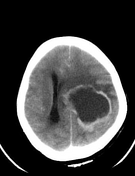
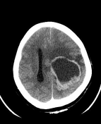
Non-contrast
large intra-axial space-occupying lesion with peripheral hyperdensity and central hypodensity in the left hemisphere (fronto-parietal region, extending into corona radiata and internal capsule)
with surrounding edema and mass effect - compressing left lateral ventricle, effacement of sulcal spaces and midline shift (about 14mm) to right
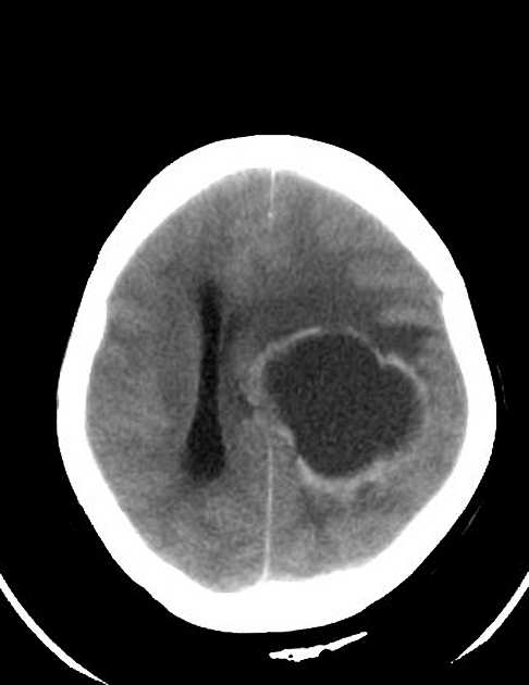
Post-contrast and delayed
shows peripheral enhancement
Case Discussion
This patient had a history of Breast cancer treated surgically with post-op, chemoradiotherapy. Imaging features are consistent with cerebral metastases with peripheral hemorrhage and central necrotic area in a known case of breast cancer.


 Unable to process the form. Check for errors and try again.