
Presentation
Low back pain.
Patient Data
Age: 80 years
From the case:
Schmorl node (annotated MRI)


Download
Info
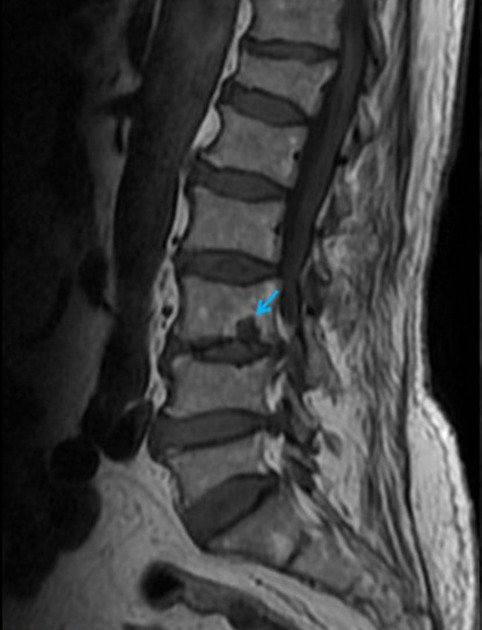
Both T1-WI and T2-WI show a well-defined hypo-intense lesion in the lower endplate of L3 (arrow). The lesion is connected with the L3-L4 intervertebral disc.
Case Discussion
Schmorl nodes represent vertical disk prolapses through areas of weakness in the vertebral endplate. Schmorl nodes are often multiple and occur predominantly in the thoracolumbar spine. Recently formed Schmorl nodes can be painful and may be indistinguishable from inflammatory or tumoral disease in terms of signal intensity. The identification of endplate defects or intranuclear cleft bending of the disk at either CT or MR imaging is helpful in making the correct diagnosis of acute Schmorl nodes.


 Unable to process the form. Check for errors and try again.