
Patient Data
Age: 25 years
Gender: Female


Download
Info

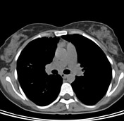
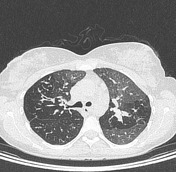
CT shows bilateral cylindrical bronchiectasis, predominant in the lower, lingula and middle lobes associated with peribronchial thickening and some mucous plugs, especially in the lower lobes. This finding is accompanied by centrilobular nodules with tree in bud morphology, compatible with inflammatory disease of distal airway.
Case Discussion
Extensive bilateral cylindrical bronchiectasis, centrilobular nodules associated with tree in bud morphology, inflammatory findings consistent with distal airway compromise .


 Unable to process the form. Check for errors and try again.