
Presentation
Abdominal pain
Patient Data
Age: 19 months old
Gender: Female
From the case:
Intussusception
Download
Info
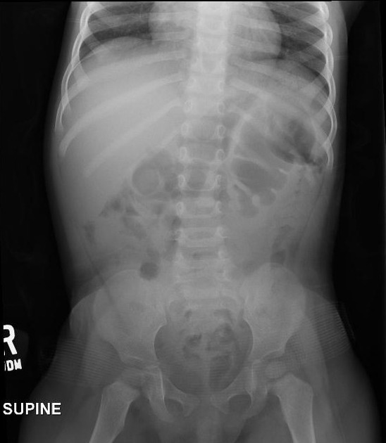
Soft tissue mass projects over the right lower abdomen. No bowel dilation.
From the case:
Intussusception
Download
Info
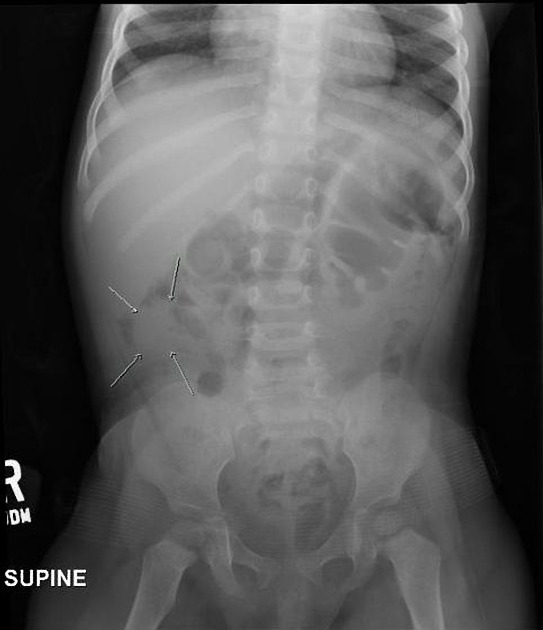
Arrows pointing toward soft tissue mass within right lower quadrant.
From the case:
Intussusception
Download
Info

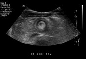
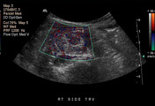
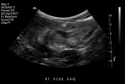
Transverse US shows concentric alternating echogenic and hypoechoic bowel walls keeping with target sign of Intussusception. Found to be of ileocolic type.
Case Discussion
The original abdominal radiograph was read negative. With high suspicion for intussusception, an ultrasound was performed. The intussusception was within the right lower abdomen, and in retrospect likely represents the soft tissue density on the abdominal radiograph as described above.


 Unable to process the form. Check for errors and try again.