
Presentation
Increasing SOB ?infection.
Patient Data
Age: 40 years
Gender: Female
From the case:
Thoracic lymphadenopathy - breast cancer (chest x-ray)
Download
Info
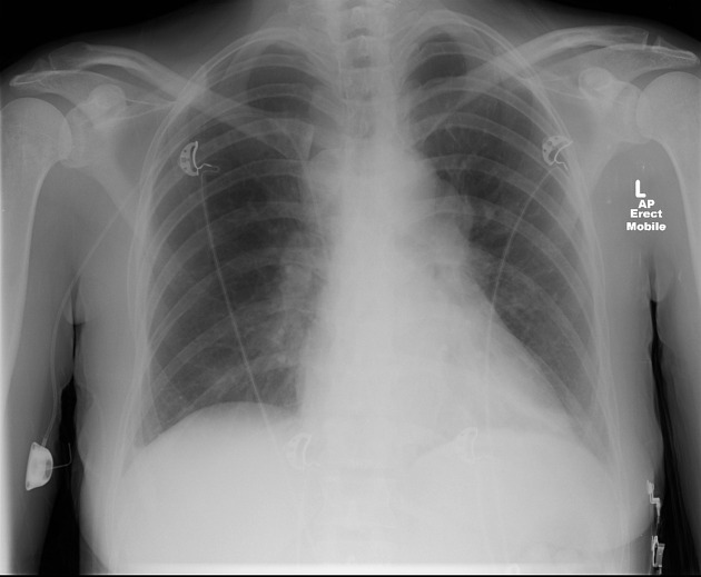
No focal consolidation, collapse or pneumothorax. No large pleural effusion. Widened right paratracheal stripe, loss of normal AP window contour and left hilar mass, most in keeping with lymphadenopathy. Right sided brachial port with tip projecting at the cavo-atrial junction.
From the case:
Thoracic lymphadenopathy - breast cancer (chest x-ray)
Download
Info
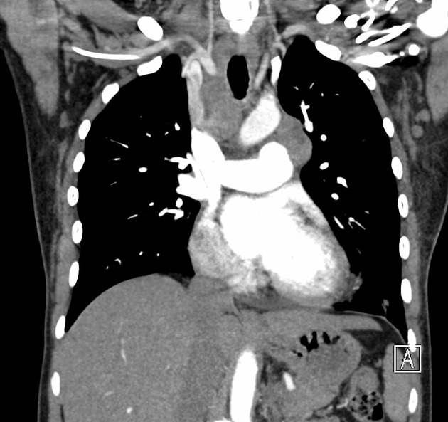
CT confirms right paratracheal and AP window lymphadenopathy.
Case Discussion
The patient has a known history of breast cancer and this lymphadenopathy represents metastatic disease.


 Unable to process the form. Check for errors and try again.