
Presentation
Pre-operative chest x-ray
Patient Data

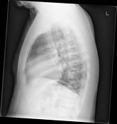

Frontal and lateral chest radiographs reveals mildly enlarged cardiac silhouette, H-shaped vertebrae, bilateral patchy humeral head sclerosis, absent splenic shadow, calcified gallstones in the RUQ abdomen, and an enlarged hepatic silhouette. No acute pulmonary pathology evident.
Case Discussion
Frontal and lateral chest radiographs demonstrates multiple bone and soft tissue findings associated with sickle cell disease:
H-shaped vertebrae with central end plate depression involving the superior and inferior endplates
bilateral patchy humeral head sclerosis consistent with epiphyseal infarction
absent splenic shadow with loops of bowel replacing the left upper quadrant abdomen suggesting autosplenectomy. Later confirmed on CT (not shown)
calcified gallstones seen in the right upper quadrant abdomen from radio-opaque bilirubin/pigmented gallstones
enlarged heart secondary to chronic anemia
enlarged liver shadow (not completely imaged) in keeping with hepatomegaly


 Unable to process the form. Check for errors and try again.