
Presentation
Indeterminate pleural nodule.
Patient Data
Age: 64
Gender: Male

Download
Info

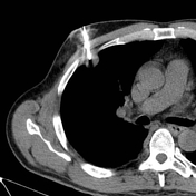
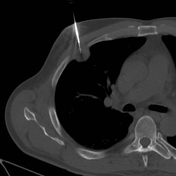
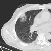
CT images show a co-axial needle into the pleural nodule. After the procedure, control images show a small local pneumothorax and an intra-alveolar hemorrhage seen as ground-glass infiltrates in local lung parenchyma.
Case Discussion
The advanced position of the co-axial needle into the pleural nodule is the most likely cause of a possible lung punching by the core-biopsy needle.
Parenchymal hemorrhage is a possible complication of thoracic biopsy and is typically self-limited. Hemoptysis can be the presenting symptom, as was the case in this patient.


 Unable to process the form. Check for errors and try again.