
Presentation
Fever and shortness of breath
Patient Data

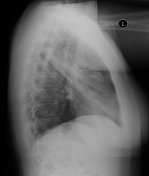

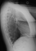
On the PA radiograph there is opacity with air bronchograms in the left midzone with loss of visualization of the left heart border. The appearance is consistent with consolidation within the left upper lobe. This is confirmed on the lateral image.
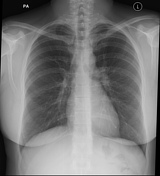

Chest radiograph 3 months later shows resolution of the consolidation with re-appearance of the left heart border.
Case Discussion
Left upper lobe pneumonia with a good example of the silhouette sign on the PA radiograph helping to localize the mid-zone opacity to the left upper lobe. The fact that visualization of the left heart border is lost implies that the pulmonary consolidation must directly abut the heart, which therefore places it towards the anterior of the chest within the lingula portion of the left upper lobe rather than within the more posteriorly located lower lobe.


 Unable to process the form. Check for errors and try again.