
Presentation
Syncope and dyspnea.
Patient Data

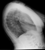
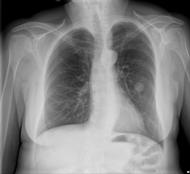
The 2 cm nodule in the left upper lobe is stable compared to the previous CXR from 5 years earlier. No appreciable calcification or fat is seen. Lungs and pleural spaces are otherwise clear. The cardio-mediastinal contour is normal.


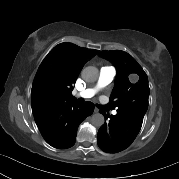
There is diagnostic contrast opacification of the pulmonary arteries. There is no pulmonary embolus. No signs of right heart strain.
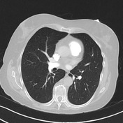
There is a well defined, slightly heterogenous solitary pulmonary nodule located anteriorly in the left upper lobe measuring 21 x 20 mm. This nodule splays the surrounding pulmonary vessels. It has a density of -10 HU with a mixed pattern of soft tissue and microscopic fat. No calcification.
There are linear densities seen in both bases in keeping with bibasal atelectasis. The remaining lungs and pleural spaces are clear.
There is no lymph node enlargement or other mediastinal abnormality.
No bony or soft tissue abnormality seen.
Conclusion
- No pulmonary embolus.
- Solitary pulmonary nodule in the left upper lobe that has not changed over the last 5 years with a low (negative) density representing a pulmonary hamartoma (a benign lesion).


 Unable to process the form. Check for errors and try again.