
Presentation
Presented to A&E with non-specific symptomatology and weight loss. No fever.
Patient Data
At the initial presentation, a chest x-ray was taken (unavailable) showing diffuse miliary nodules.


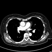
1.5 cm spiculated mass in the apical segment of the right lower lobe.
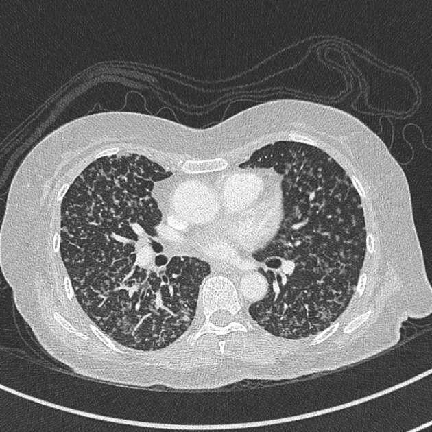
Innumerable miliary nodules, with a rather ground-glass appearance throughout both lungs.
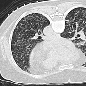

Diffuse miliary nodules throughout the lungs.
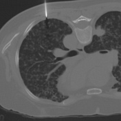
1.5cm spiculated lesion not targeted due to its adherance to the left oblique fissure and deep location due to higher pneumothorax risk.
A tiny procedural pneumothorax is evident.
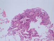
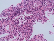
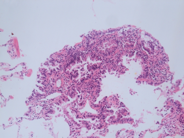
Lung biopsy
Neoplastic cuboidal to columnar cells growing along alveolar walls in a lepidic fashion.
Positive for CK7 and TTF1.
Appearances are those of a non-mucinous bronchoalveolar carcinoma of the lung
Case Discussion
Traditionally and still often referred to as bronchoalveolar carcinoma (BAC), this form of lung cancer is now classified as adenocarcinoma in situ, minimally invasive adenocarcinoma and invasive adenocarcinoma of the lung based on histological findings.
Classically, it presents insidiously with an unresolving pneumonic process, but can also present as a pulmonary nodule or cluster of diffuse nodules as in this case.
Three recognized radiographic patterns are described:
- single mass or nodular form: ~45%
- consolidative form: ~30%
- multinodular form: ~25%


 Unable to process the form. Check for errors and try again.