Presentation
Abdominal discomfort and swelling. VP shunt inserted previously for hydrocephalus. No fever or previous abdominal surgery.
Patient Data
![](https://prod-images-static.radiopaedia.org/images/15946645/712c3993d2acfca8a727e0512ae31b_thumb.jpeg)
![This study is a stack](/packs/stack-YQKLCKBI.gif)
![](https://prod-images-static.radiopaedia.org/images/15946663/1d701090af787641401b42b24f243d_thumb.jpeg)
![This study is a stack](/packs/stack-YQKLCKBI.gif)
![](https://prod-images-static.radiopaedia.org/images/15946645/712c3993d2acfca8a727e0512ae31b_big_gallery.jpeg)
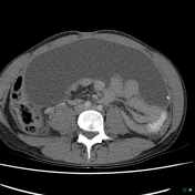
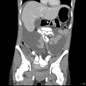
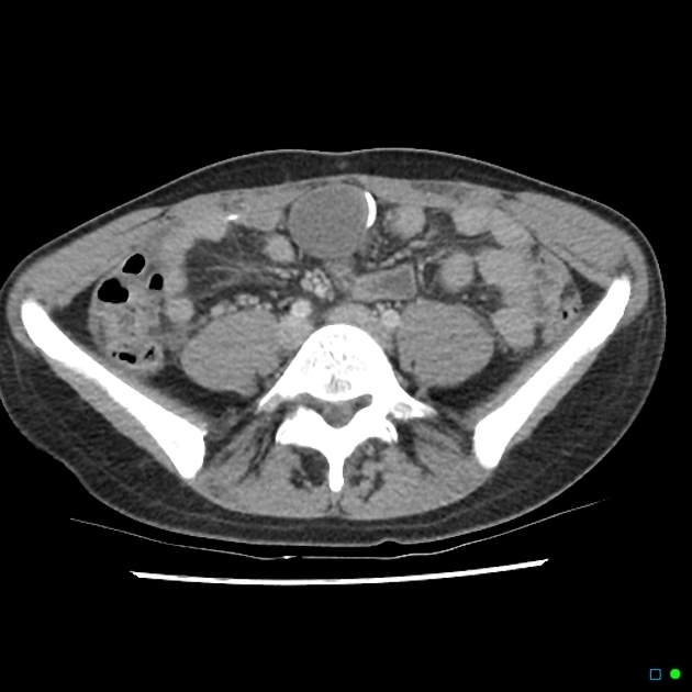
Very large well-defined fluid collection in the anterior upper abdomen associated with the peritoneal end of a VP shunt.
![](https://prod-images-static.radiopaedia.org/images/15946519/123d69d68d77c1372972c2dc894864_thumb.jpeg)
![](https://prod-images-static.radiopaedia.org/images/15946553/63fcc460b9d6064c15624d259aa6fd_thumb.jpeg)
![](https://prod-images-static.radiopaedia.org/images/15946584/37186ae44f785bfc436d070f2c61d3_thumb.jpeg)
![](https://prod-images-static.radiopaedia.org/images/15946519/123d69d68d77c1372972c2dc894864_big_gallery.jpeg)
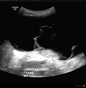
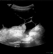
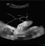
Fluid collection in the abdomen containing internal septations, but otherwise non-echogenic fluid consistent with CSF
![](https://prod-images-static.radiopaedia.org/images/15946514/e5765a94f7624a560f2d72983edac2_thumb.jpeg)
![This study is a stack](/packs/stack-YQKLCKBI.gif)
![](https://prod-images-static.radiopaedia.org/images/15946517/c64456c7ee0b34062d5eae978b82ce_thumb.jpeg)
![This study is a stack](/packs/stack-YQKLCKBI.gif)
![](https://prod-images-static.radiopaedia.org/images/15946514/e5765a94f7624a560f2d72983edac2_big_gallery.jpeg)
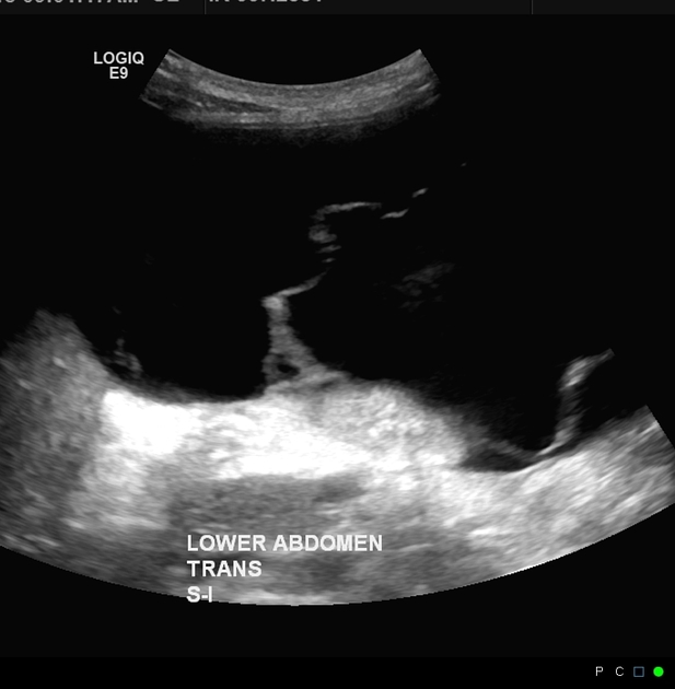
Almost complete resolution following drainage and replacement of the drainage tube.
![](https://prod-images-static.radiopaedia.org/images/15946369/0ead9647b013e1a0609284204d3b64_big_gallery.jpeg)
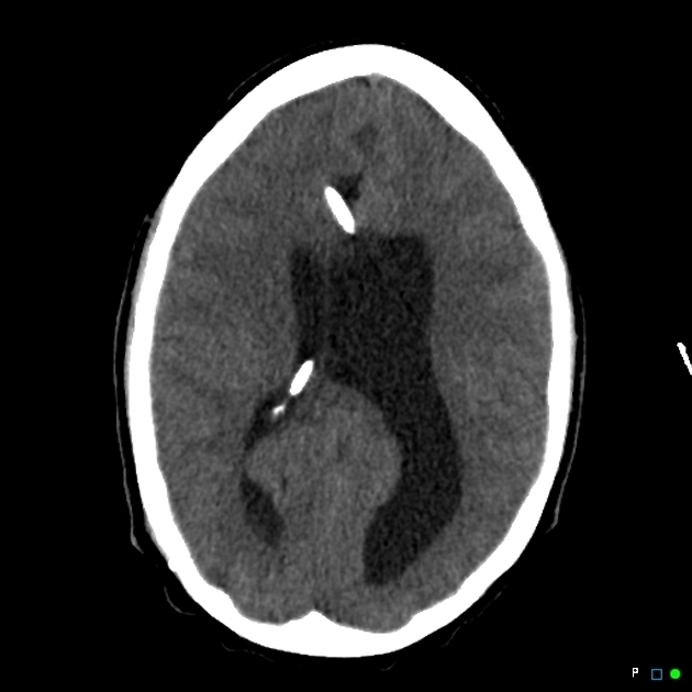
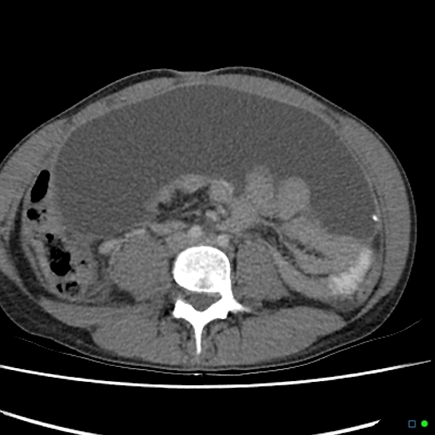
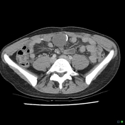
CT brain showing 2 shunt tubes in situ, the anterior one not connected to the peritoneum.
Case Discussion
Loculated peritoneal CSF "pseudocysts" - so-called as they have a fibrous wall rather than epithelial are rarely associated with VP shunts. As they are slowly expanding, they can reach enormous sizes before presentation. They are thought to be due to low-grade infection or peritoneal adhesions. Many are idiopathic.