
Presentation
An infant with no past medical history. On physical exam, the left side of the limbs is larger than the right.
Patient Data
Note: This case has been tagged as "legacy" as it no longer meets image preparation and/or other case publication guidelines.

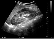
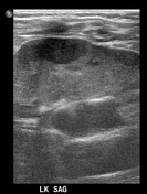
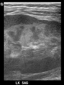
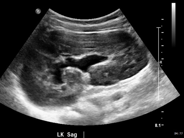
The left kidney is larger and demonstrates lobularity with hypoechoic cortical nodularity. Differential at the times is persistent fetal lobularity versus nephrogenic rest.


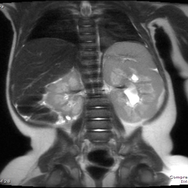
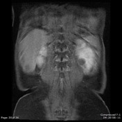
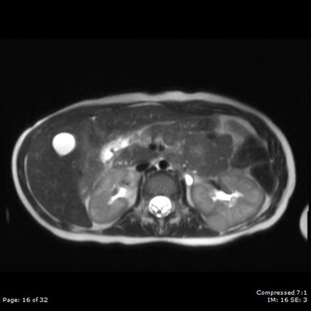
Coronal and axial T2 HASTE shows multiple nodular lesions of varying sizes and shapes in bilateral kidneys, left greater than right, predominantly peripherally located, with features most consistent with bilateral nephroblastomatosis.
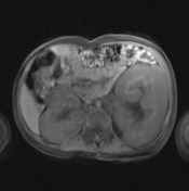
Contrast MRI at the time of presentation shows no discrete heterogeneity or area of focally abnormal contrast enhancement identified, as to convincingly suggest malignant transformation.

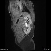
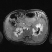
Pre and Post-contrast MRI at a follow-up study 1 year from the initial presentation which demonstrates multiple hypointense nodules in both kidneys have increased in size as described above. Left lower pole lesion shows faint internal and peripheral enhancement. Findings are concerning for malignant transformation of the previously identified perilobar nephrogenic rests.
Case Discussion
Nephrogenic rest (NR): persistent metanephric blastema 1
in the periphery: perilobular
within the kidney: intralobular
multiple NRs: nephroblastomatosis
NRs are important to diagnose and follow because they are precursors to Wilms tumor.
In some studies, up to 35% of diffuse hyperplastic perilobar NR transform to Wilms Tumor. This is considered the most at risk group
The patient, in this case, demonstrates this in the initial ultrasound images.
Other syndromes associated with NRs (and Wilms tumor) are
sporadic aniridia
On imaging, the NRs appear homogeneous on all imaging modalities. This is a very important distinction to Wilms tumor, which is heterogeneous. The NRs do not enhance to the same degree as the surrounding kidney
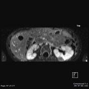
The patient, in this case, demonstrates homogeneous enhancement less that kidney parenchyma on the initial MRI images.
Unfortunately, a repeat MRI one year later demonstrates increased peripheral and internal enhancement, findings concerning for malignant transformation.


 Unable to process the form. Check for errors and try again.