
Presentation
Decreased attention with parkinsonism features.
Patient Data


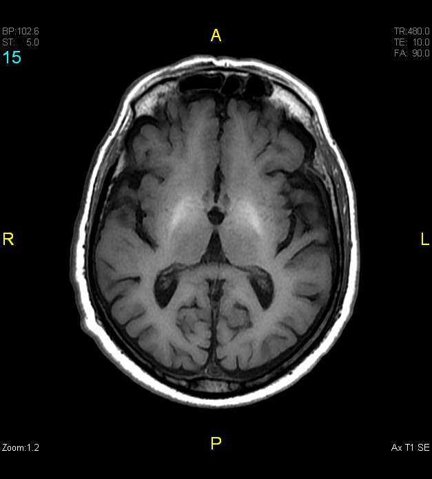
Symmetric T1 hyperintense signals are seen arising from the globi pallidi, ventrolateral thalami, and substantia nigra of the midbrain. There is sparing of the cerebellum, red nucleus, and putamen. No features of midbrain atrophy. Age proportionate cerebral atrophic changes.

Cirrhotic liver identified in upper abdominal slices.
Case Discussion
Following detection of the symmetric T1 hyperintense affliction of globus pallidus, ventrolateral thalami and substantia nigra, a possibility of hepatocerebral degeneration was considered. Previous imaging of the patient was reviewed. A CT chest study done a month ago revealed the cirrhotic nature of liver.
Acquired hepatocerebral degeneration is an often underdiagnosed condition which can present with parkinsonism features. This also highlights the importance of not ignoring the T1 weighted image of brain, which by convention has always been a source of 'anatomical' information. One of those rare instances when pathology is only identified in this sequence.


 Unable to process the form. Check for errors and try again.