
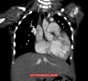
Presentation
Child with cardiomegaly and shortness of breath.
Patient Data
Age: 1 year
Gender: Female
From the case:
Cor triatriatum


Download
Info
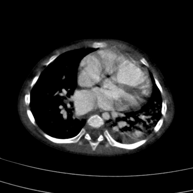
CT demonstrates a membrane dividing left atrium into a proximal chamber, containing the pulmonary venous confluence, and a distal "true" left atrium, which contains the left atrial appendage.
An anomalous vein drains the atelectatic left upper lobe of the lung and joins the left brachiocephalic vein.
The pulmonary arteries, right ventricle and right atrium are dilated.
From the case:
Cor triatriatum
Download
Info
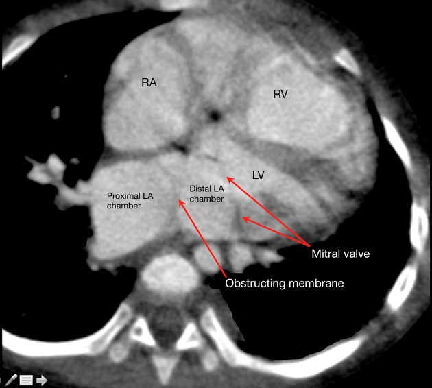
Annotated CT image showing an obstructing fibromuscular membrane separating the left atrium into proximal and distal chambers.
Case Discussion
This is an intraoperatively confirmed case of cor triatriatum with partial anomalous pulmonary venous return (left upper lobe).


 Unable to process the form. Check for errors and try again.