
Presentation
Abdominal mass, flank pain and gross hematuria.
Patient Data
Age: 75 years
Gender: Male
From the case:
Renal cell carcinoma


Download
Info
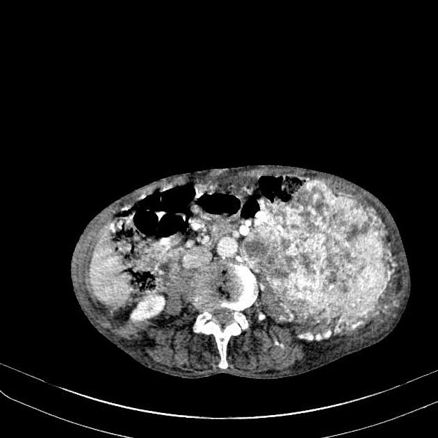
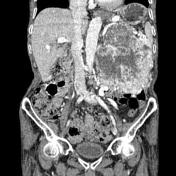
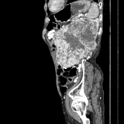
There is a large enhancing mass arising from the left kidney. The mass shows central necrotic areas as well as increased vascularity. The mass invades the abdominal wall.
Case Discussion
Renal cell carcinomas (RCC) are primary malignant adenocarcinomas derived from the renal tubular epithelium and are the most common malignant renal tumor. They usually occur in 50-70 year old patients and macroscopic hematuria occurs in 60% of the cases.
On imaging they have a variety of radiographic appearances, from solid and relatively homogeneous to markedly heterogeneous with areas of necrosis, cystic change and hemorrhage.


 Unable to process the form. Check for errors and try again.