
Presentation
Diffuse abdominal pain and leukocytosis
Patient Data

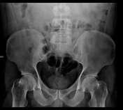
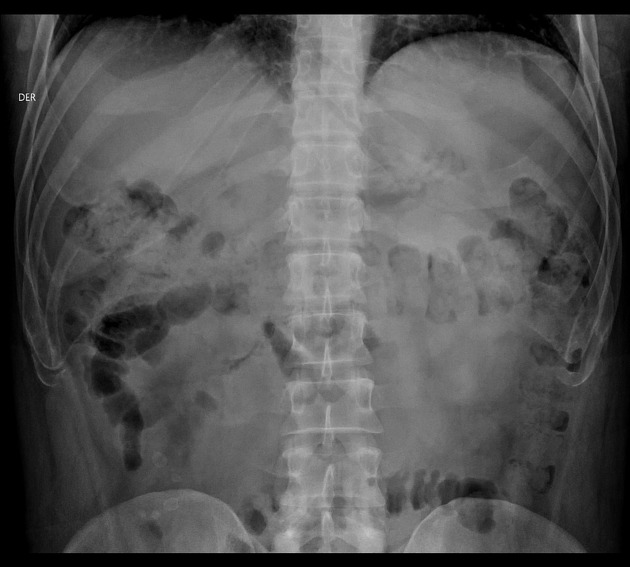
Abdominal AP X-ray demonstrate the presence of five appendicolith projecting over the right iliac crest. Ultrasonography was inconclusive because the appendix was difficult to evaluate due to the presence of abundant intestinal gas.
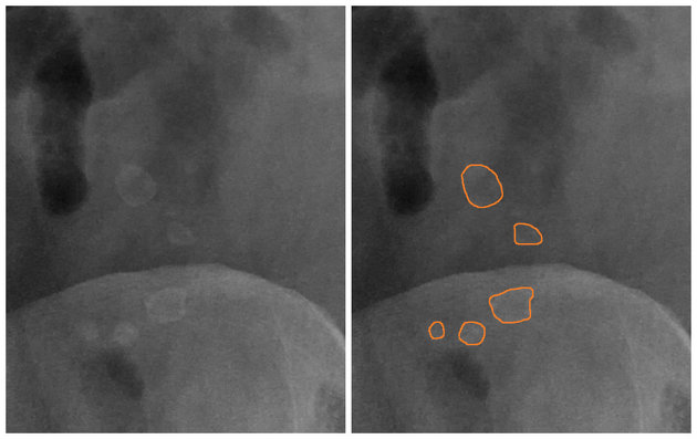
Annotated image shows the presence of five appendicoliths.
The patient underwent open surgery after X-ray findings and acute appendicitis was found.
Case Discussion
The appendicolith, also known as “fecolith” or “corpolith”, represents calcified deposits in the appendix, and contributes to the pathogenesis of acute appendicitis. It is defined as an area of high attenuation measuring ≤ 1 cm that is located in the pericecal areas, or in cases of perforation in the Morrison's (Douglas) pouch.
The finding of an appendicolith may be sufficient evidence to perform a prophylactic appendectomy in asymptomatic patients, given the higher rate of perforation at the time of acute appendicitis.


 Unable to process the form. Check for errors and try again.