
Presentation
Weight loss and cough. TB?
Patient Data
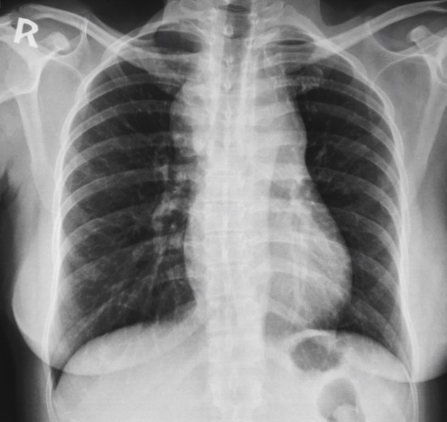
Near symmetrical upper anterior mediastinal mass. Left sided hilar overlay sign.
Widened companion shadow overlying the left clavicle.
Heart size normal. Lungs clear.


CT CHEST
Large anterior mediastinal mass with bulk dimensions of 7.2 x 5cm. Lungs clear. Bilateral lymph node enlargement at the root of the neck.
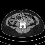
CT ABDOMEN
Extensive para-aortic, celiac axis, porta hepatis and iliac lymph nodes measuring up to 2.3 cm. Solid organs normal. No focal bone lesion.
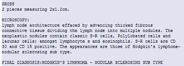
Excision Bx of a neck node undertaken
Reports in keeping radiological suspicious of lymphoma.
Case Discussion
A good case for long case or viva of a fellowship examination.
Start with the plain film to assess:
- the candidate's approach to a common film
- their understanding of how to determine mediastinal compartment the pathology lies in
- knowledge to give a reasonable differential diagnosis
- to 'earn' the CT to give a more confident definitive diagnosis
- question on the best method of ascertaining a tissue diagnosis
Additional discussion of US-guided neck node biopsy vs surgical excision vs CT-guided mediastinal mass biopsy.


 Unable to process the form. Check for errors and try again.