
Presentation
Chronic knee pain and recent fall 2 weeks ago.
Patient Data

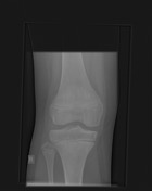
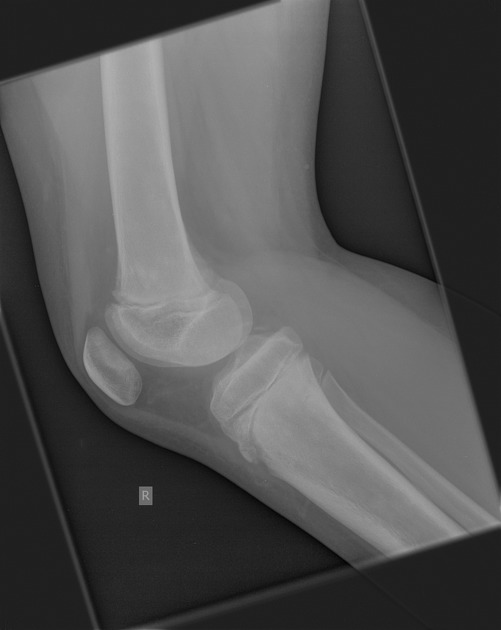
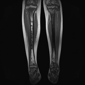
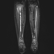
Subtle, patchy sclerotic changes in the distal femoral and proximal tibial metaphyses.


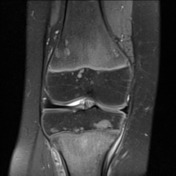
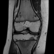
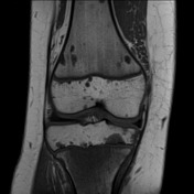
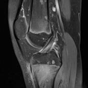
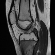
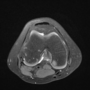
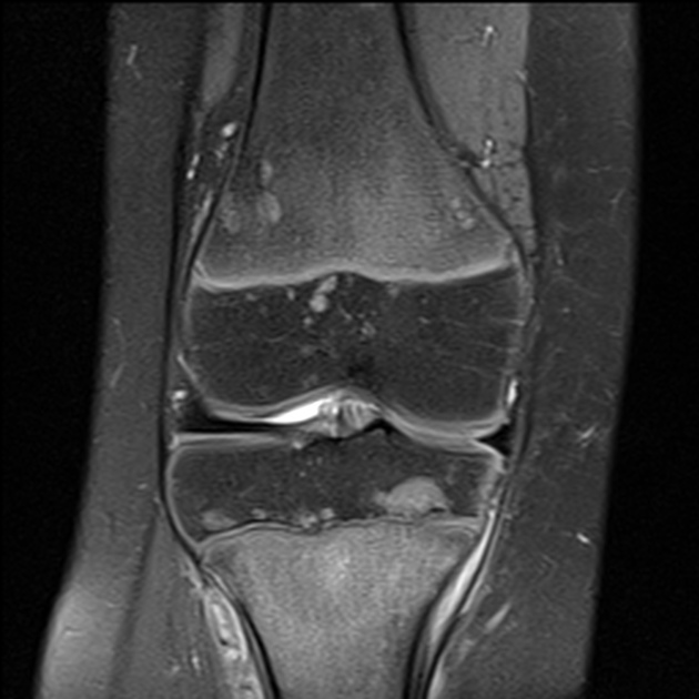
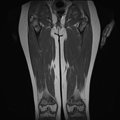
Numerous well circumscribed intramedullary lesions of variable size are demonstrated throughout the imaged periarticular femur, tibia, fibula and patella, involving both the metadiaphyses and epiphyses.
The lesions are of low T1 signal. Most are of high signal on the PD fat sat (fluid sensitive) sequences, and low signal on the PD acquisition. There is minimal cortical scalloping, but no overt lytic process.
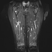
Additionally, there is more confluent fluid signal within the anterior tibial metaphysis, with suspected anteromedial microtrabecular fracture.
The cruciate and collateral ligaments, menisci and chondral surfaces are intact.
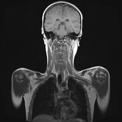
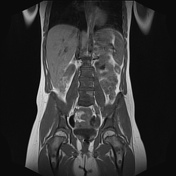
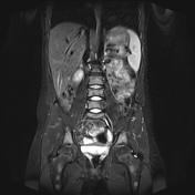
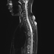
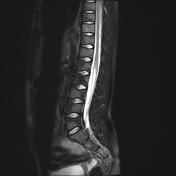

Subsequent whole body MRI has shown extensive, patchy marrow infiltration of the entire skeleton.
Case Discussion
This is pathologically proven acute lymphoblastic leukemia (ALL).
The initial diagnosis is confounded by the fairly atypical, patchy and well defined bone lesions - rather than diffuse marrow infiltration - in a clinically well child, presenting with a single region complaint (knee pain) in an outpatient setting. Extensive metastases is a differential.
Concurrent anterior tibial marrow edema with suspected microtrabecular fracture and overlying pretibial edema, most likely due to acute injury, leading to presentation and ultimately her diagnosis.


 Unable to process the form. Check for errors and try again.