
Presentation
Long-standing Parkinson disease
Patient Data
Age: 65 years
Gender: Male
From the case:
Displaced deep brain stimulators
Download
Info

- implantable pulse generator (IPG) in right chest wall
- ventriculoperitoneal shunt (VPS) coursing vertically on left
- tracheostomy cannula
- left-sided pleural effusion
From the case:
Displaced deep brain stimulators

Download
Info
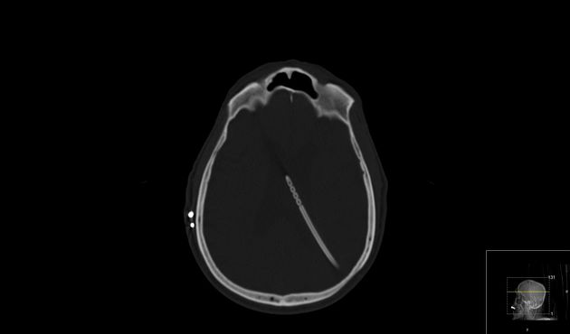
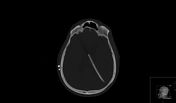
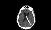
DBS electrode pair seen tunneling subcutaneously along right parietal bone, not reaching burr holes at coronal sutures. Tracts through brain are evident. VP shunt in left ventricle.
Case Discussion
The patient had been suffering from Parkinson disease for over 35 years, with eventual vocal cord paralysis. Several years prior to the above radiographic studies, he had deep brain stimulators implanted, but to no avail.


 Unable to process the form. Check for errors and try again.