
Presentation
Left abdominal pain
Patient Data
The only available previous radiologic examination was an upright PA chest radiograph:
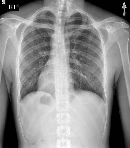
Situs inversus: right-sided heart and aorta; gastric bubble and spleen on right, liver on left.
Since the chest radiograph had been obtained several years earlier, there was no way of ascertaining proper patient positioning by the radiographer.
Situs inversus was verified at ultrasonography:

Unpaired abdominal organs on contralateral side, IVC left of aorta.
Appendix identified in left lower abdomen, measuring 10 mm in diameter, non-compressible, with periappendiceal fat stranding.
Case Discussion
A 40-year-old male presented to the ED complaining of left abdominal pain. Lab results were notable for leukocytosis with left shift and an elevated CRP.
The only examination documented in the PACS was an 'old' chest radiograph, which clearly showed situs inversus. There was no way of ascertaining that the patient had been properly positioned for said radiograph, though, so situs inversus was verified on ultrasonography.
Subsequently, the appendix was sought and found in the left lower abdomen, showing signs of acute inflammation.
Surely enough, an inflamed appendix was excised laparoscopically.
Left lower abdominal pain in a person with situs inversus should raise the suspicion of acute appendicitis, as all the organs are 'flipped' horizontally.


 Unable to process the form. Check for errors and try again.