
The appendix or vermiform appendix (plural: appendices) is a blind muscular tube arising from the cecum, the first part of the large bowel.
On this page:
Gross anatomy
The appendix arises from the posteromedial surface of the cecum, approximately 2-3 cm inferior to the ileocecal valve, where the three longitudinal bands of the taeniae coli converge. It is a blind diverticulum which is highly variable in length, ranging between 2 and 20 cm. The appendiceal mesentery is called the mesoappendix 1,2.
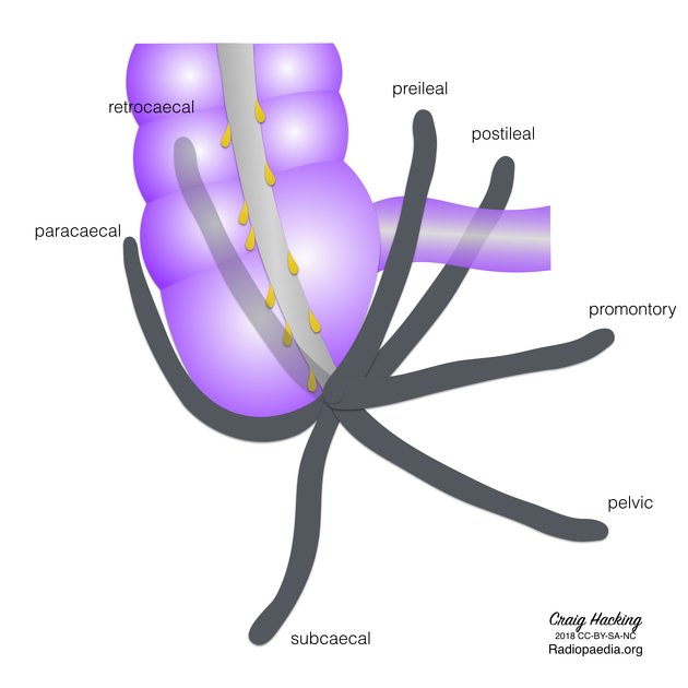
The tip of the appendix can have a variable position within the abdominal cavity 1,2:
retrocecal (65-70%)
pelvic (25-30%)
pre- or post-ileal (5%)
promontory
paracaecal
subcecal
The appendix is also attached to the ileocecal junction by the ileocecal fold (bloodless fold of Treves). The ileocecal fold is a peritoneal structure which runs from the antimesenteric aspect of the ileum, is reflected over the ileocecal junction, and joins the base of the mesoappendix. A fossa termed the inferior ileocecal recess is between the ileocecal fold and mesoappendix.
Arterial supply
appendicular artery, an end artery arising from the ileocolic artery (itself from the superior mesenteric artery) 1
Venous drainage
similarly named veins draining to the portal venous system
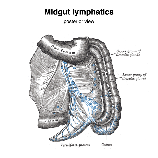
Lymphatic drainage
the appendix is rich in lymphoid tissue which peaks around 20 years of age
see article: cecum
Innervation
see article: cecum
Variant anatomy
additional arterial supply from accessory appendicular artery
duplex appendix: very rare
horseshoe appendix: extremely rare
Radiographic features
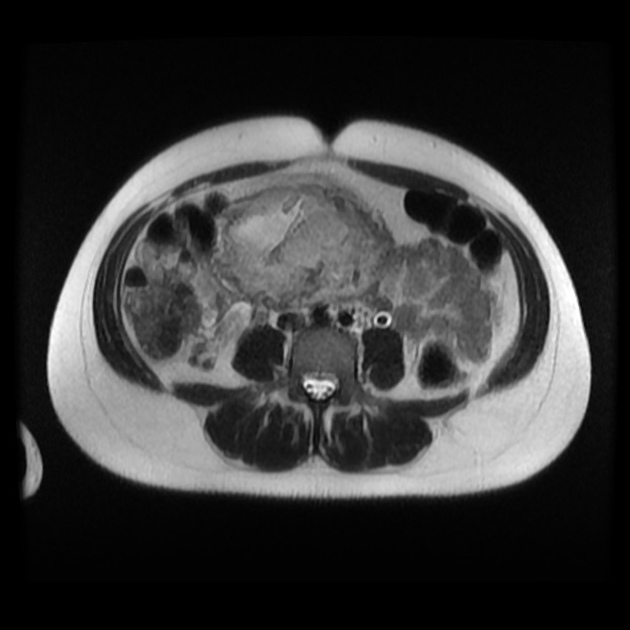
The normal appendix can be identified most of the time without a significant difference in detection rate across the following modalities 5:
ultrasound: ~70%
MRI: ~70%
CT: ~85%
Plain radiograph
an appendicolith is seen in 10% of patients, with 90% going on to develop appendicitis 1
the appendix can fill with contrast during a barium enema study
Ultrasound
A dynamic ultrasound technique using a sequential 3-step patient positioning protocol can increase the visualization rate of the appendix 3. In the study, patients were initially examined in the conventional supine position, followed by the left posterior oblique (LPO) position (45° LPO) and then a “second-look” supine position. Reported detection rates increased from 30% in the initial supine position to 44% in the LPO position and a further increase to 53% with the “second-look” supine position. The authors suggested that the effect of the LPO positioning step improved the acoustic window by shifting bowel contents.
History and etymology
'Vermis' is the Latin word for worm, hence vermiform means worm-like. The word appendix originates from the Latin verb 'appendere' meaning "to hang from".


 Unable to process the form. Check for errors and try again.