
Presentation
Referred from the opthalmologist with ? right orbital vascular malformation.
Patient Data


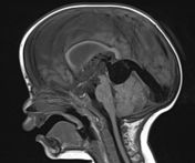
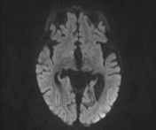
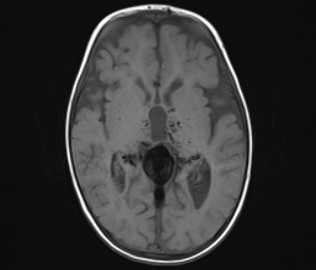
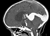
Aneurysmal dilated midline venous pouch is noted representing dilated vein of Galen measuring 2.5 x 3.5 cm in maximum diameters. It is joining a normally situating straight sinus which is dilated measuring 1.2 cm, joining dilated torcular herophili from which extend extensively dilated straight and sigmoid sinuses.
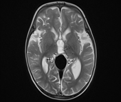
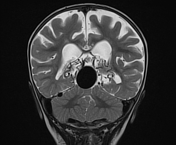
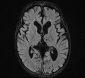
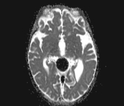
Mild to moderate dilation of the supratentorial ventricles secondary to venous hypertension. Superior sagittal sinus is patent receiving extensively dilated cortical veins.
Bilateral prominent peri-optic nerve CSF spaces (Peri-optic hydrops) secondary to venous hypertension. Normal CT appearance of the eye globes, extra-ocular muscles, lacrimal glands, retrobulbar regions and peri-orbital soft tissues. No intra or extra-conal lesions could be detected. Normal dimensions of the optic canals. Intact orbital bony frame-works.

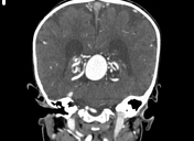
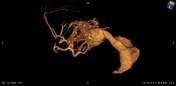
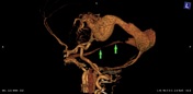
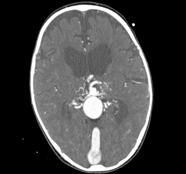
Bilateral dilated tortuous cisternal tuft of the vessels are noted in quadrigeminal cistern and tela choroidae on both sides made of dilated arteries and early draining veins in keeping with AVM likely made by posterior choroidal vessels, thalmo-perforators, and peri-chalosal arteries from PCAs (Choroidal AVMs).
Extensively dilated right superior petrosal sinus extending from right sigmoid sinus joining cavernous sinus with secondary asymmetrical extensive dilation and venous congestion of the right cavernous sinus draining the markedly dilated superior and inferior ophthalmic veins. The ophthalmic veins are joining dilated anterior facial vein, pre-nasal and pre-malar vein as well as retro mandibular vein.
Case Discussion
This case represents imaging features of choroidal type of vein of Galen malformation (VOGM) with extensive intracranial venous hypertension reflected by mild to moderate Hydrocephalic changes and bilateral optic nerve hydrops. Right cavernous sinus venous congestion is noted drained by dilated right superior petrosal sinus with secondary orbital venous congestion explaining clinical presentation of exophthalmos.
Choroidal type of VOGM represents multiple feeders including thalamoperforating, choroidal and pericallosal arteries located in the subarachnoid space in the choroidal fissure. It usually presents in the neonatal period and progresses to high output cardiac failure.


 Unable to process the form. Check for errors and try again.