
Presentation
Progressive leg weakness and sensory changes.
Patient Data

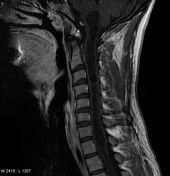
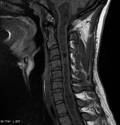
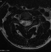

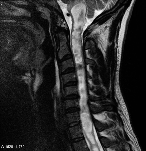
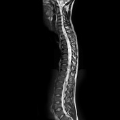
The cervical cord is markedly expanded by cystic dilatation, presumably representing a very prominent syrinx. Areas of signal loss on T2 weighted images likely represent flow/pulsation artefact, rather than blood products. No solid or enhancing component can be identified.

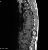
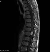
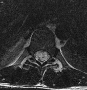
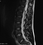
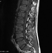
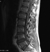
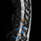
In the lower thoracic cord is a vividly enhancing nodule with associated prominent serpiginous flow voids both within the mass and over the surface of the cord, and among the cauda equina. It is an isolated abnormality, associated with marked expansion of the entire cord by a large syrinx.
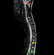
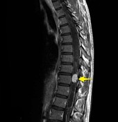
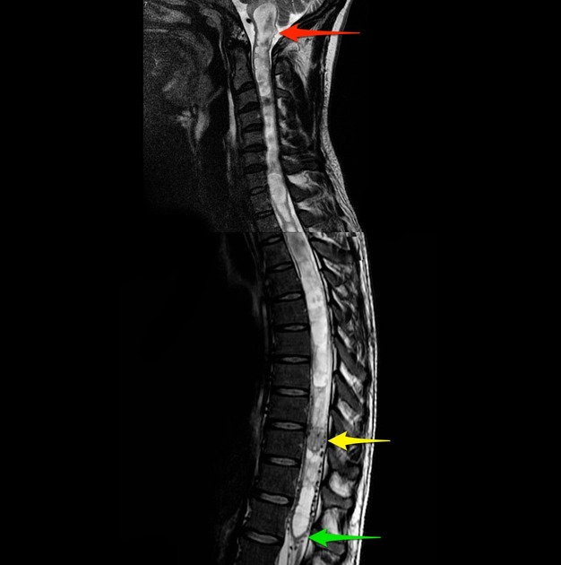
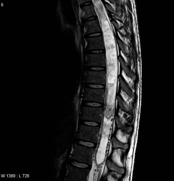
An intramedullary nodule (yellow arrow) which demonstrates very bright contrast enhancement and is associated with holocord syrinx ( * ) extending above and below the lesion from the medulla (red arrow) to the tip of the conus (green arrow).
Numerous enlarged flow voids are noted both within the cord (orange arrow) and on the surface of the cord (blue arrows).
Case Discussion
Histology proven hemangioblastoma of the spine. Note the large flow voids and marked holocord syringomyelia.


 Unable to process the form. Check for errors and try again.