
Presentation
Headaches, seizures, developmental delay. MRI examination performed for the first time.
Patient Data
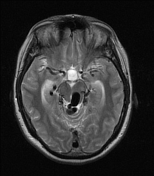

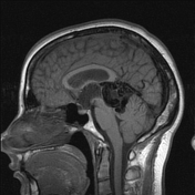
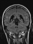
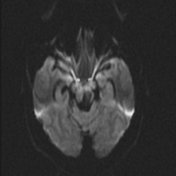

Vein of Galen aneurysmal malformation, compressing the Sylvian aqueduct.
Lateral and 3rd ventricles are moderately dilated without transependymal edema. Deep cerebral veins, vein of Rosenthal, and superior cerebellar veins are dilated bilaterally.
Convexity subarachnoid space narrowing.
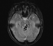
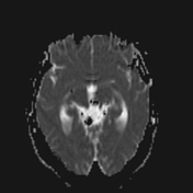
No diffusion restriction. Signal loss within the malformed vein of Galen seen on DWI due to T2 blackout effect; there is a mixture of dark and bright pixels even on the corresponding ADC map.
Dural venous sinuses seem unremarkable.
No T2* blooming.
Case Discussion
It was a big surprise to reveal the vein of Galen malformation causing obstructive hydrocephalus in a 30-year-old patient. Such a diagnosis in adults is extremely rare and if left untreated it is associated with poor clinical outcome, with a reported 76.7% mortality rate.


 Unable to process the form. Check for errors and try again.