
Presentation
This patient is known to have SVC obstruction, underwent SVC recannulization, which was thought to be complicated by injury.
Patient Data
Age: 35 year old
Gender: Male


Download
Info
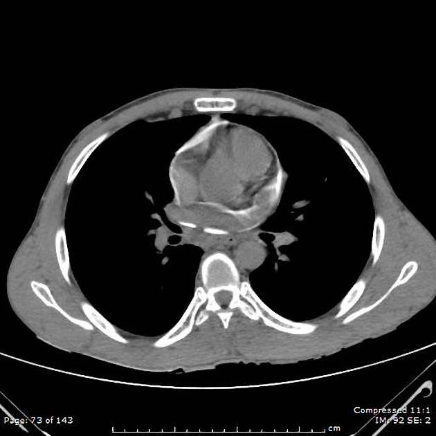
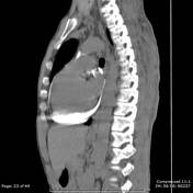
Contrast media seen in the pericardial space due to complicated attempt for recanlization of the SVC.
Case Discussion
The patient had a complicated SVC obstruction recannulization, and injury of the SVC was suspected.
CT scan with IV contrast was done which showed contrast media pooling in the pericardial sac confirming the presumed SVC injury.


 Unable to process the form. Check for errors and try again.