
Presentation
Pleuritic upper back pain. SOB. On immunosupression. ?Infection
Patient Data
Age: 65 years
Gender: Male
From the case:
Multifocal lung adenocarcinoma


Download
Info
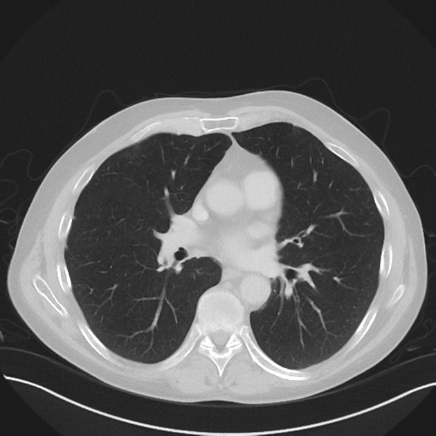
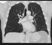
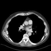
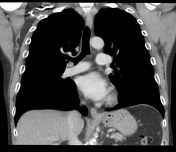
Multiple bilateral lung lesions including:
- multiple ground glass nodules in both upper lobes
- left upper lobe cavitating nodule measuring 18 mm
- right upper lobe subpleural bubbly lesion with peripheral ground glass change measuring 33 mm
- solid lesion in apical segment left lower lobe
Mild background emphysema. No endobronchial or endotracheal lesions. No significantly sized axillary, hilar or mediastinal lymphadenopathy. No pericardial or pleural effusion. Moderate coronary artery vascular calcification. No large cardiac vegetations.
Case Discussion
The differential on this CT includes multifocal adenocarcinoma, vasculitis, metastases and cavitating infection. On repeat CT 3 weeks later there had been no change. A lung biopsy was performed targeting the left upper lobe lesion. This confirmed primary lung adenocarcinoma. The other lesions are therefore most likely adenocarcinoma.


 Unable to process the form. Check for errors and try again.