
Presentation
Worsening abdominal distension. Concern for malignacy.
Patient Data
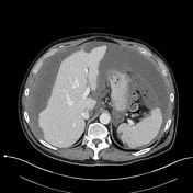

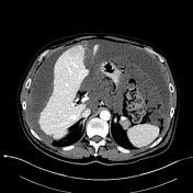
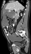

Diffuse, extensive peritoneal disease. Extensive scalloping and deformity of the liver and spleen with low attenuation material. Thick omental caking throughout the abdomen up to 4.7 cm anterior-posterior in the mid abdomen. Numerous sites of serosal implants including stomach, large bowel, and small bowel. Multiple areas of peritoneal nodularity and thickening. No small bowel obstruction is identified.
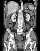
There are a few small calcifications with possible mass or implants in the region of the appendix in the right lower quadrant.
PATHOLOGY REPORT (edited):
Final pathology diagnosis from omental biopsy: Mucinous and low-grade neoplastic gastrointestinal type epithelium.
The abundant mucin and scant low-grade neoplastic gastrointestinal type epithelium are consistent with pseudomyxoma peritonei.
Case Discussion
In this case, the extensive scalloping of the liver and spleen suggest rupture of a mucinous neoplasm resulting in pseudomyxoma peritonei, rather than broadly stating there is peritoneal carcinomatosis.
A close look in the right lower quadrant reveals a few calcifications with an irregular appendix or implants, suggesting ruptured appendiceal mucinous adenocarcinoma as the cause (which also happens to be the most common cause of this condition).
Because pathologic diagnosis is ultimately required, it is appropriate to guide the ordering physician toward omental biopsy, which is very safe and can be performed under ultrasound.


 Unable to process the form. Check for errors and try again.