
Presentation
Thigh mass for 2 years. Now getting much bigger. No weight loss. Lipoma or sarcoma?
Patient Data
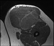

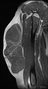
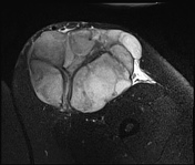
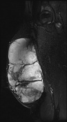
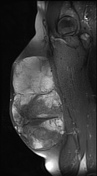

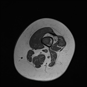
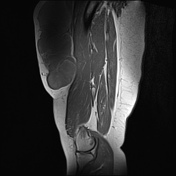
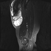
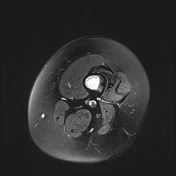
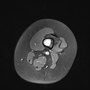
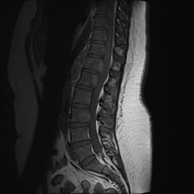
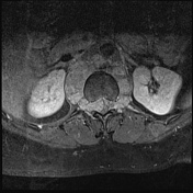
Well-delineated 20.4 x 14.7 x 11 cm lobulated mass in the thigh abutting, but not infiltrating the thigh musculature.
The mass contains multiple thin septae and fatty elements. Mild enhancement of the solid components.
No local lymphadenopathy.
Multiple high T2 foci within the femur, the largest 6cm at the proximal metaphysis.
Comment: appearances highly suggestive of a liposarcoma
The signal changes in the femur suggest osseous involvement.
Recommend;
1. MRI full femur length to include knee and hips joints
2. CT CAP

Patient recalled to ensure full upper leg covered
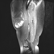
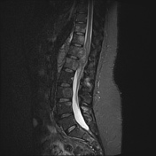
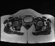
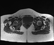
Distal femur covered. Whole femur covered in conjunction with the original imaging.
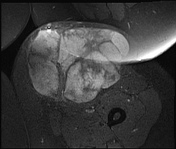
Huge thigh mass as previously reported in keeping with a liposarcoma.
Multiple infiltrative marrow lesions in the whole length of the femur. The largest 6.1 cm in the distal diaphysis and the most distal lesion of all in the metaphysis measuring 1.4 cm.
Comment: liposarcoma with osseous femoral skip lesions.


Chest:
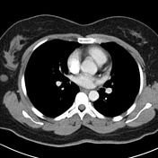
4 mm subpleural ground glass nodules in the lateral segment of the left lower lobe.
1.5 cm lesion in the upper outer aspect of the right breast and lower inner aspect of the right breast.
No mediastinal nodes.
Abdomen:
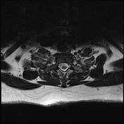
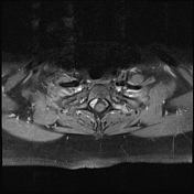
Circumferential rind of paravertebral soft tissue measuring 2.5 cm in depth at T12-L1. The soft tissue extends into the epidural/dural spaces at these levels.
Minor scalloping of the posterior aspects of T12 and T1 likely related to the aforementioned soft tissue extension.
Three metastases the largest measuring 2.6 and 1.6 cm in segment 5 of the liver.
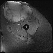
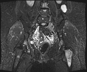
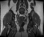
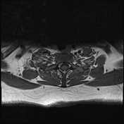
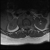
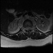
6.2 cm mass in the right side of the pelvis compressing the lateral bladder wall. 3.5 cm peritoneal metastases posterolateral the left kidney.
Comment:
1. T12-L1 paraspinal tissue with soft tissue extension - advise MRI whole spine to assess
2. liver and pelvic lesions - consistent with metastases in the context of the thigh mass (liposarcoma)
3. right breast lesions - dedicated imaging merited

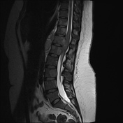
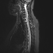
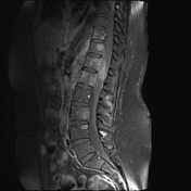
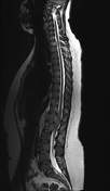
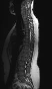
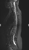
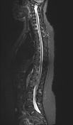
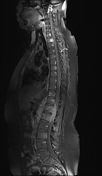
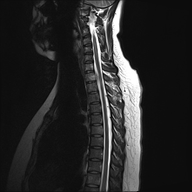
Counting from C2, the last unfused vertebra is taken as L5. Spinal cord ends normal at L1 level.
Moderately enhancing masses are involving almost the entire spine, hypointense on T1 and intermediate signal on T2 images, though preserved vertebral body and disc heights.
There is associated paraspinal extension of the lobulated masses with anterior epidural extension at T11–L2 levels, resulting in significant spinal canal stenosis and compression of the cord and nerve roots in the cauda equina.
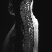
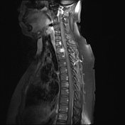
Anterior epidural extension of the mass is also noted at C7–T2 levels causing mild to moderate spinal canal stenosis and abutting the cord.
6.7 cm lobulated soft tissue mass in the right side of pelvic cavity, compressing right lateral wall of the bladder. 3.5 cm left retroperitoneal nodule.
Case Discussion
The breast and thigh lesion were both biopsied.
THIGH: A malignant tumor composed of lipoblast in a myxoid stroma.
Conclusion: mxyoid liposarcoma
BREAST: round to short spindle cells in a myxoid background. Vimentin positive. S100, CD68, CD34 and desmin negative.
Conclusion: myxoid liposarcoma
Quite a remarkable case of diffuse metastatic disease from a thigh liposarcoma including the rare occurance of breast metastases.
The thigh mass was present for several years prior and presumably metamorphasized into a malignancy.


 Unable to process the form. Check for errors and try again.