Presentation
Neck mass. Biopsy and chemotherapy. Follow up scan. Clinically now a normal examination.
Patient Data
![](https://prod-images-static.radiopaedia.org/images/40684478/302d895b81645a2cab87667f9df656_thumb.jpeg)
![This study is a stack](/packs/stack-YQKLCKBI.gif)
![](https://prod-images-static.radiopaedia.org/images/40684607/46667582f0b7664fcb0a5ad8043b8a_thumb.jpeg)
![This study is a stack](/packs/stack-YQKLCKBI.gif)
![](https://prod-images-static.radiopaedia.org/images/40684844/65ab89bfc538005fd8c57c005b4629_thumb.jpeg)
![This study is a stack](/packs/stack-YQKLCKBI.gif)
![](https://prod-images-static.radiopaedia.org/images/40684702/44c8c496433ab42affe726a053bccd_thumb.jpeg)
![This study is a stack](/packs/stack-YQKLCKBI.gif)
![](https://prod-images-static.radiopaedia.org/images/40684965/86913530f34a0509c7105fd007bc68_thumb.jpeg)
![This study is a stack](/packs/stack-YQKLCKBI.gif)
![](https://prod-images-static.radiopaedia.org/images/40685054/e142ee46f4d9f9db1c5f388eedc5d2_thumb.jpeg)
![This study is a stack](/packs/stack-YQKLCKBI.gif)
![](https://prod-images-static.radiopaedia.org/images/40685143/8e418fcf3d45b75f4407ed5adebb18_thumb.jpeg)
![This study is a stack](/packs/stack-YQKLCKBI.gif)
![](https://prod-images-static.radiopaedia.org/images/40685231/318c0ee7277d9b5501e09113ce9898_thumb.jpeg)
![This study is a stack](/packs/stack-YQKLCKBI.gif)
![](https://prod-images-static.radiopaedia.org/images/40685346/ac8c9fc9e113688ec6c459247d09f1_thumb.jpeg)
![This study is a stack](/packs/stack-YQKLCKBI.gif)
![](https://prod-images-static.radiopaedia.org/images/40685449/f2b6143ece214975c42488101943c8_thumb.jpeg)
![This study is a stack](/packs/stack-YQKLCKBI.gif)
![](https://prod-images-static.radiopaedia.org/images/40685735/ea34f431e80d75a0e565984cc1fb36_thumb.jpeg)
![This study is a stack](/packs/stack-YQKLCKBI.gif)
![](https://prod-images-static.radiopaedia.org/images/40684478/302d895b81645a2cab87667f9df656_big_gallery.jpeg)
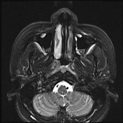
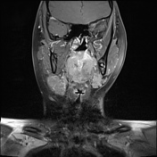
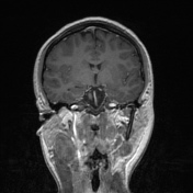
Asymmetrical soft tissue in the right nasopharynx obliterating the fossa of Rosenmuller. No parapharyngeal extension.
Bilateral retropharyngeal adenopathy measuring 1.8cm on the right and 1.7cm
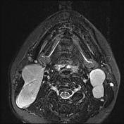
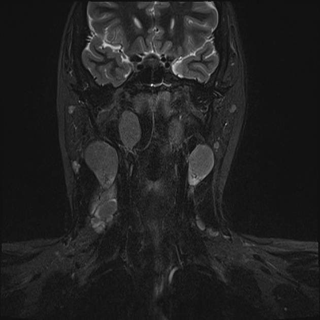
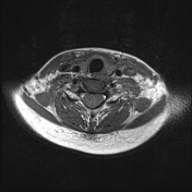
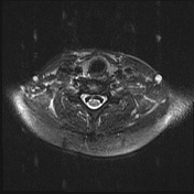
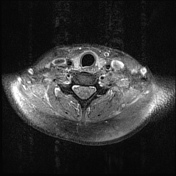
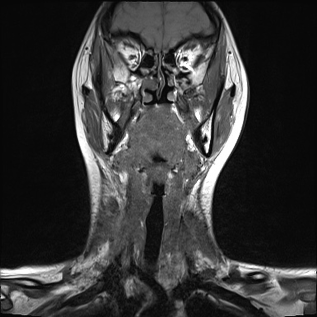
Bilateral large level II cervical lymphadenopathy, the largest measuring 4.2cm on the right and 2.7cm on the left and level III lymphadenopathy measuring up to 1.8cm and right level IV lymphadenopathy measuring 2cm.
![](https://prod-images-static.radiopaedia.org/images/40438461/24c5baec43b96b866cd0f953300e32_thumb.jpeg)
![This study is a stack](/packs/stack-YQKLCKBI.gif)
![](https://prod-images-static.radiopaedia.org/images/40438332/478cde8e530e4ea38a8765ee53b919_thumb.jpeg)
![This study is a stack](/packs/stack-YQKLCKBI.gif)
![](https://prod-images-static.radiopaedia.org/images/40438647/6331f210beb57024f8e52cc81a50dd_thumb.jpeg)
![This study is a stack](/packs/stack-YQKLCKBI.gif)
![](https://prod-images-static.radiopaedia.org/images/40438560/7bc6fded3196c08b2caf855d84fe6a_thumb.jpeg)
![This study is a stack](/packs/stack-YQKLCKBI.gif)
![](https://prod-images-static.radiopaedia.org/images/40438798/d16147e6796c23969647b3cc5656db_thumb.jpeg)
![This study is a stack](/packs/stack-YQKLCKBI.gif)
![](https://prod-images-static.radiopaedia.org/images/40438736/0c31b5f8b149d9bfc72fda57eb2ea3_thumb.jpeg)
![This study is a stack](/packs/stack-YQKLCKBI.gif)
![](https://prod-images-static.radiopaedia.org/images/40438876/66ea8a6a898c8857aa1bd530c65a90_thumb.jpeg)
![This study is a stack](/packs/stack-YQKLCKBI.gif)
![](https://prod-images-static.radiopaedia.org/images/40438971/a719b26d9934f9d7a32522eb5e8c19_thumb.jpeg)
![This study is a stack](/packs/stack-YQKLCKBI.gif)
![](https://prod-images-static.radiopaedia.org/images/40439065/2a98cef87d98a8badb87f2a035e6ce_thumb.jpeg)
![This study is a stack](/packs/stack-YQKLCKBI.gif)
![](https://prod-images-static.radiopaedia.org/images/40439353/cabda3237b6f6d77034a77821df288_thumb.jpeg)
![This study is a stack](/packs/stack-YQKLCKBI.gif)
![](https://prod-images-static.radiopaedia.org/images/40438461/24c5baec43b96b866cd0f953300e32_big_gallery.jpeg)
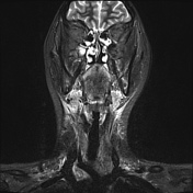
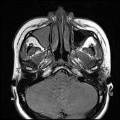
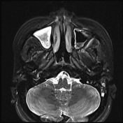
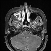
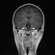
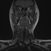
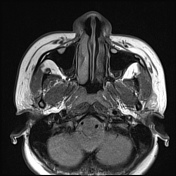
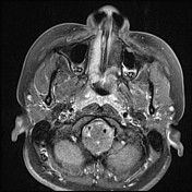
The right nasopharyngeal mass is no longer evident. Symmetry of the soft tissues of the nasopharynx.
The bilateral retropharyngeal lymphadenopathy has resolved.
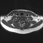
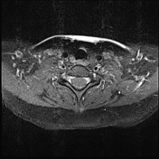
The extensive bilateral cervical chain lymphadenopathy has regressed in its entirety.
Reactive 9mm right submandibular node.
Fluid in the right maxillary and frontal sinuses. Left mastoid air cells contain fluid.
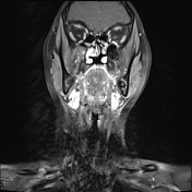
Comment: Complete regression of right nasopharyngeal mass and metastatic lymphadenopathy
Case Discussion
This young patient presented with a right-sided neck mass. Ultrasound-guided biopsy confirmed a nasopharyngeal nodal metastases and a subsequent ENT assessment and biopsy confirmed this as the primary. It is not uncommon to see nodal disease large in volume compared to the nasopharyngeal primary as in this case.
Radiologically the nasopharyngeal carcinoma was T1,N3a,M0.
Chemotherapy was undertaken with the follow-up MRI one year later demonstrates complete radiological regression of the previous extension of metastatic nodal disease and the primary nasopharyngeal tumor.