
Presentation
Intermittent hematuria and hemoptysis. No arthralgia. Anti-MPO and Anti-PR3 postive.
Patient Data


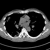
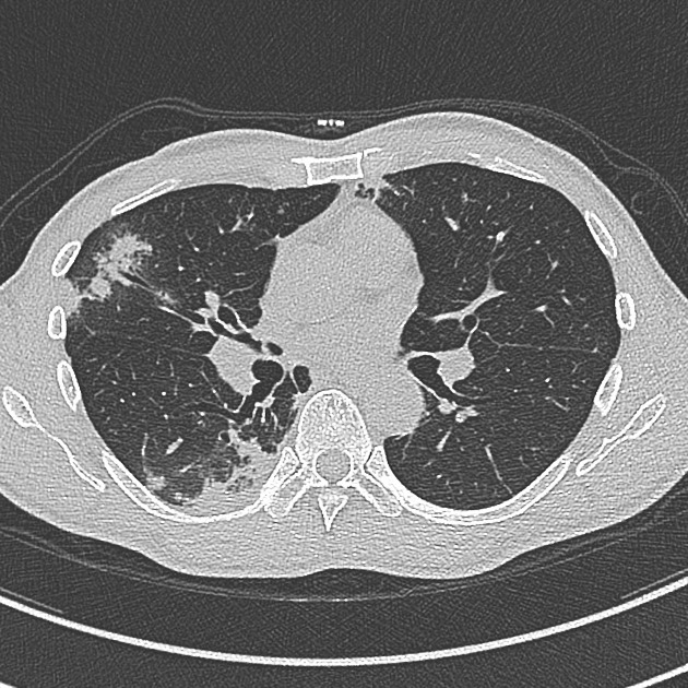
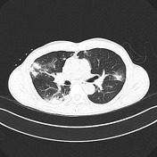
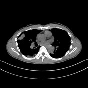
Multiple focal areas of consolidation in both lungs measuring upto 2 cm, most of which are subpleural in location. Several are wedge shaped and have a small amount of surrounding groundglass opacification.
Minor right apical bronchiectasis.
No endobronchial lesion.
No mediastinal lymphadenopathy.

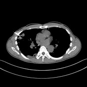
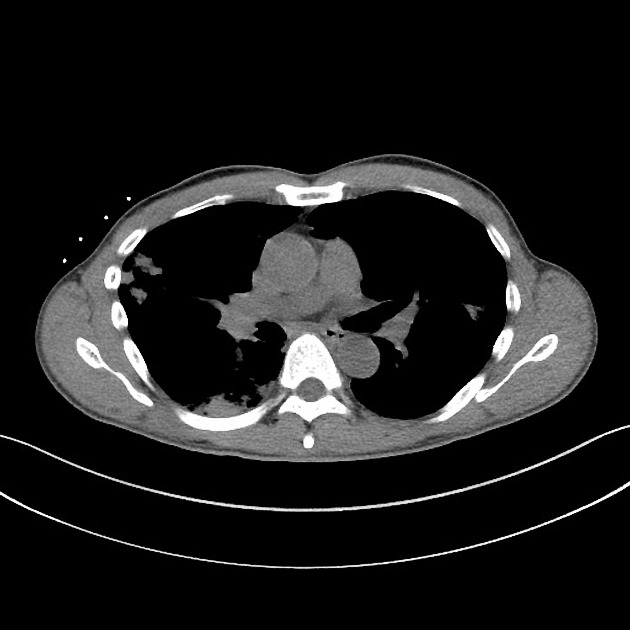
Right upper lobe subpleural 1.4 cm lesion targeted.
20 G core biopsy x 2 taken.
The patient has hemoptysis immediately after the biopsy. Several clots (1-3 mL).
3 mm post procedural pneumothorax.
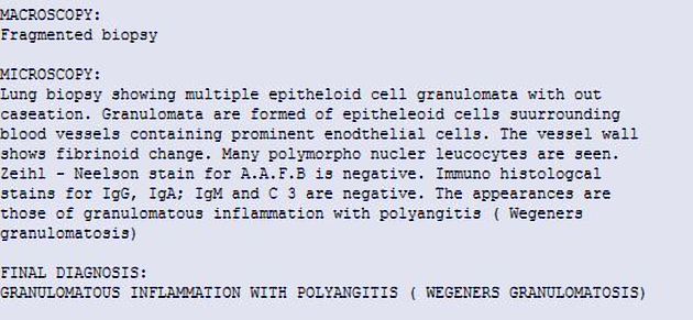
LUNG BIOPSY RESULT: Granulomatous inflammation with polyangiitis
Case Discussion
Granulomatosis with polyangiitis (GPA) is a multisystem necrotizing non-caseating granulomatous vasculitis involving the small to medium-sized arteries, capillaries, and veins. It most commonly affects the respiratory system and kidneys as in this case.
This patient also had laboratory evidence of renal impairment and a renal biopsy identified concurrent renal involvement.
Four types of pulmonary involvement are recognized, one of which is typified by peripheral wedge-like consolidation as illustrated in this case.
The post lung biopsy hemoptysis self-resolved. Immunosuppressive treatment was commenced.


 Unable to process the form. Check for errors and try again.