
Presentation
Head injury from a plastic sheet impact
Patient Data
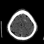

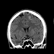
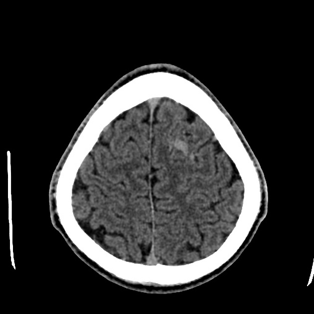
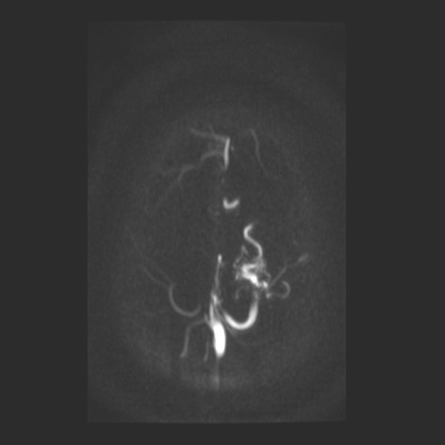
Tuft of serpiginous vessels seen in left superior frontal gyral surface.
No intracranial bleed was identified. So I opted to do a correlative MR Venogram to identify if it is anything more than a developmental venous anomaly.
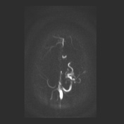
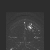
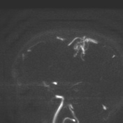
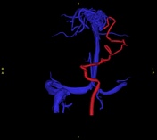
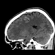
Incidentally detected arteriovenous malformation involving left superior frontal lobe with nidus of approximately 2.1 x 2.0 cm, with arterial feeders from cortical branches of left middle cerebral artery and venous drainage into superior sagittal sinus.
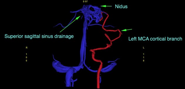
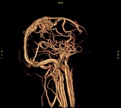
Annotated depiction of the arterial feeder, nidus and venous drainage.
Case Discussion
Imagine the luck of this boy. He had an incidental head injury. No post injury symptoms, but anxious parents said let's do a head CT trauma protocol. CT technician flagged left frontal subarachnoid hemorrhage. I reviewed the images and immediately realized this is an abnormal vessel. Let's just do a venogram. And that revealed this tuft of vessels having arterial feeders from cortical branches of the left middle cerebral artery.


 Unable to process the form. Check for errors and try again.