Presentation
6-year history of cough, hemoptysis, dyspnea and wheeze. Generally 1 episode of hemoptysis per day and about 10 ml per episode.
Patient Data
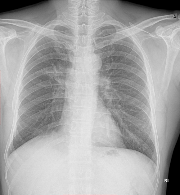
Ill-defined increased opacity in the right parahilar lung.
No other significant abnormality.
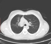

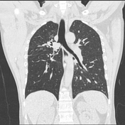

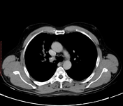

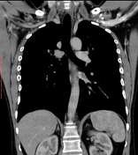

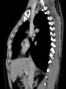

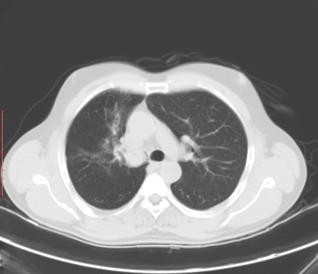
A well-defined endoluminal homogenously enhancing soft tissue attenuation lesion in the right main bronchus extending proximally towards the carina, laterally into the origin of right upper lobe bronchus and inferiorly into the bronchus intermedius causing right upper lobe bronchial luminal compromise with adjacent lung changes in the form of atelectatic strands and ground glass opacities involving the apical and anterior segments of the right upper lobe.
Single focus of calcification.
Case Discussion
Histopathologically proven case of bronchial carcinoid associated with punctuate calcification and bronchial stenosis with distal mild atelectasis and obstructive pneumonitis. Marked contrast enhancement is common due to vascularity and can mimic pulmonary varix or pulmonary artery aneurysm.
CT is valuable to look for mediastinal extension and to reveal mucoid impaction
Bronchial fibroepithelial polyp is a differential diagnosis.


 Unable to process the form. Check for errors and try again.