
Presentation
Four days abdominal pain, loose stools and fever. Severe RIF tenderness. C-reactive protein is 31mg/L.
Patient Data


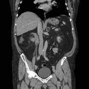
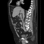
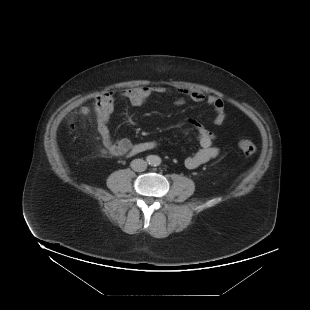
Several diverticula of the medial aspect of the appendix which are markedly inflamed with periappendiceal fat stranding. Appendix itself looks unremarkable.
No fluid collections or free gas. Several reactive lymph nodes within the draining mesentery.
Mild uncomplicated diverticulosis in the sigmoid is also noted.
Conclusion:
Appearances were suspicious for acute diverticulitis of the appendix.
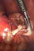
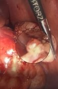
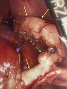

Photos from the operative field during the laparoscopic appendectomy. The diathermy forceps grasp the vermiform appendix. The diverticulum can be seen arising from the superior margin of the appendix on the first photo and magnified on the second.
Key to third photo:
- yellow arrows: vermiform appendix
- green arrow: diverticulum arising from the appendix
- blue arrows: cecum
At surgery, there were inflamed diverticula but the appendix itself was not inflamed.
Histology
Macroscopic description:
Pot labeled appendix
Appendix measuring 93 mm in length and 8 mm in diameter. Part of the serosa is missing, 40 mm away from the base. Two nodular structures identified 8 mm and 5 mm. The serosa is smooth and shiny. The lumen is filled with soft brown material.
Microscopic description:
The sections of appendix show acute inflammation. Also diverticulitis is identified in the appendix. No evidence of unusual features.
Conclusion
Appendix - Acute appendicitis. Appendiceal diverticulitis.
Case Discussion
Diverticulitis of the appendix is a well-described entity in the surgical and pathological literature. It is less well-known to many radiologists. Diverticula arising from the appendix are histologically-identical to those of the remainder of the colon, i.e. they are pseudodiverticula or false diverticula as they do not involve all three layers of the bowel wall.


 Unable to process the form. Check for errors and try again.