
Presentation
Abdominal pain and progressive distension.
Patient Data
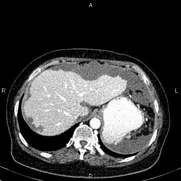
Multiple low-attenuating mass-like lesions with loculated fluid throughout the peritoneum, omentum and mesentery, accompanied by scalloping of visceral surfaces, particularly the liver. No calcification is present.
A 12 mm ill-defined hypo-attenuating lesion is noted at the superior aspect of the spleen.
A few simple cortical cysts are seen in the kidneys.
A 60 mm midline fascial defect is noted at the abdominal wall that some omental fat and bowel loops herniated through it.
Case Discussion
The features on the CT scan and the extensive scalloping of the liver strongly suggest pseudomyxoma peritonei, a syndrome of progressive intraperitoneal accumulation of mucinous ascites related to a mucin-producing neoplasm.
A mucinous tumor of the appendix most commonly causes it. Much less commonly, mucinous colon, rectum, stomach, pancreas, and urachus tumors are implicated. There is some ongoing contention as to whether primary ovarian tumors are a frequent source in their own right or whether, in these cases, the appendix is the primary site and the ovarian lesion is metastatic.


 Unable to process the form. Check for errors and try again.