
Presentation
Head trauma. Incidental findings
Patient Data


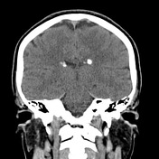
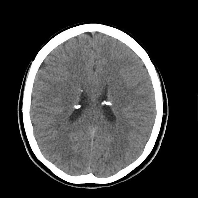
There are multiple calcified subependymal nodules along the margins of the lateral ventricles consistent with subependymal hamartomas. No parenchyma abnormality. No signs of traumatic brain injury.
CT of the brain does not show any traumatic injury but demonstrates calcified subependymal nodules (hamartomas), suspicious for tuberous sclerosis (TS).
The next step will be CT of chest and abdomen to search for other signs of TS.
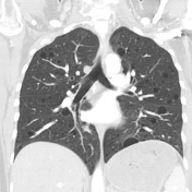


Numerous thin-walled lung cysts consistent with lymphangioleiomyomatosis are seen. No pneumothorax. No pleural effusion.
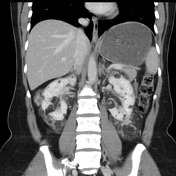


Multiple different size renal angiomyolipomas are demonstrated. There are two hypervascular lesions in the left liver lobe, best seen in arterial phase, almost isodense in venous phase, and hypodense in the delayed phase.
Case Discussion
In this case, there are three major features of tuberous sclerosis diagnostic criteria: subependymal nodules, lymphangioleiomyomatosis and renal angimyolipomas (more than 2). So patient has a definite TS complex. Interestingly, patient has no clinical symptoms or skin lesions.
Hepatic hypervascular lesions can be interpreted as angiomatous type of hepatic angiomyolipomas without macroscopic fat.


 Unable to process the form. Check for errors and try again.