
Presentation
Progredient right-sided hemiparesis. Seizures in the last few days.
Patient Data
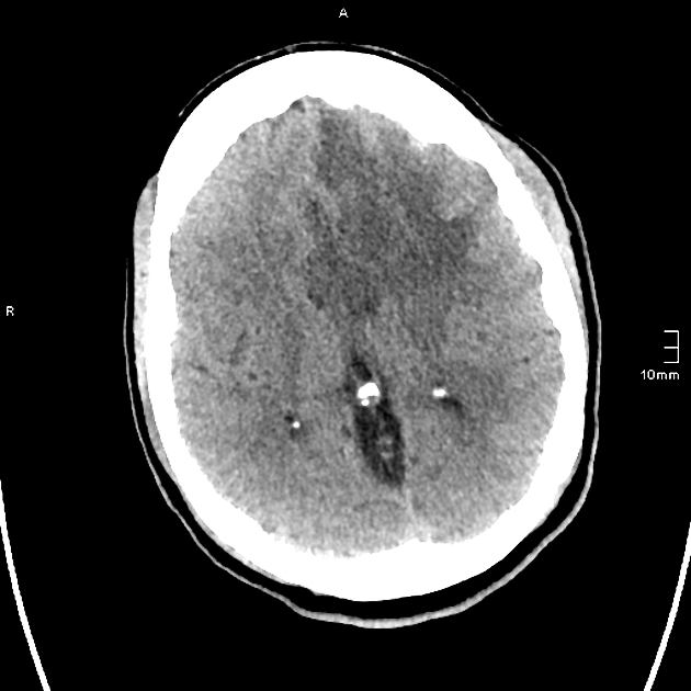
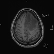
Left frontal lobe spherical mass with ring iso/hyperdensity and central hypodensity, surrounded by severe perifocal edema.
Midline shift, no herniation, no signs of hydrocephalus.
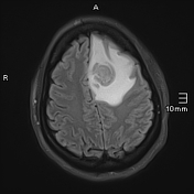


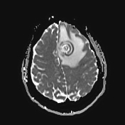
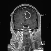

Left frontal lobe spherical mass (3 cm) with massive perifocal edema, moderate regional mass effect with effacement of the frontal sulci and left lateral ventricle.
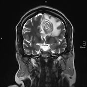
The mass shows a concentric target sign in several measurements, best seen in the T2w TSE.
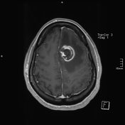
Peripheral closed ring enhancement.
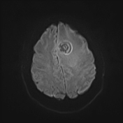
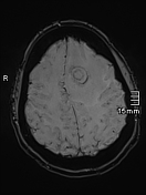
SWI shows concentric susceptibility (microhemorrhages or calcifications).
Separate Focal T2-hyperintensity in the right lentiform nucleus might be a small separate lesion.
Case Discussion
MRI findings are suspicious of an inflammatory cause.
The presence of concentric rings in the T2w images is highly suspicious for neurotoxoplasmosis.
Differential diagnoses based on imaging alone were:
1. Cerebral Toxoplasmosis in an immunocompromised patient
2. CNS lymphoma in an immunocompromised patient
3. Balo concentric sclerosis (usually showing only mild perifocal edema) in an immunocompetent patient
Blood tests revealed positive results for HIV (yet unknown to the patient).
Immunochemical findings were typical for Neurotoxoplasmosis.
Toxoplasma gondii IgG > 13 000 IU/ml
Toxoplasma gondii IgA AK 1:1024 (normal < 1:16)
HIV-1 RNA quantitatively copies/ml: >280 000
CD 4+ T-Cells 7/μl (normal 500-2000/μl)
The Patient was thus diagnosed with AIDS (Neurotoxoplasmosis being one of the AIDS-defining illnesses in an HIV infected patient) and was treated with 1. Antiretroviral therapy, 2. Antitoxoplasmosis therapy (Pyrimethamine, Clindamycin, Folate), 3. Antiepileptic medication.


 Unable to process the form. Check for errors and try again.