
Presentation
Dyspnea
Patient Data
Age: 40 years
Gender: Female
Download
Info
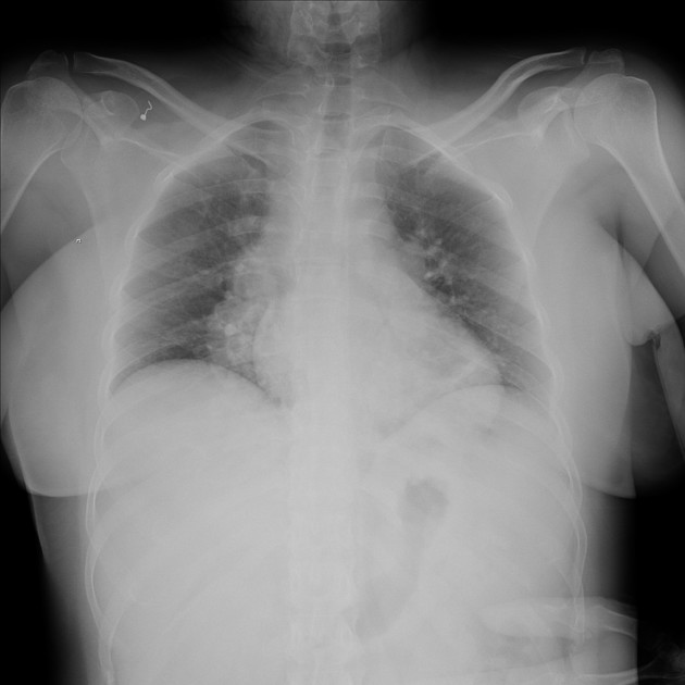
Prominent lung hila bilaterally.
Normal cardiothoracic ratio.
Clear costophrenic angles.

Download
Info
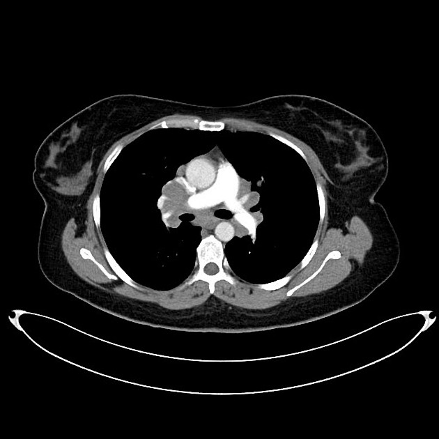
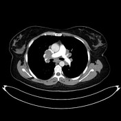
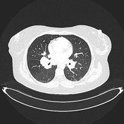
Mediastinal and hilar lymphadenopathy, lung nodules, and an enhancing hepatic nodule.
Case Discussion
A CXR was initially obtained and then CT was requested for further evaluation of prominent hila. The lung nodules were thought to be metastatic lesions.
HISTOPATHOLOGY REPORT:
Section of all specimens reveals multiple discrete non-caseating epithelioid granulomas, composed of epithelioid histiocytes and multinucleated giant cells with little residual benign lymphoid tissue in between. No acid-fast bacilli were identified by Z-N stain. No malignancy noted.
Diagnosis: Non-caseating granulomas, consistent with sarcoidosis.


 Unable to process the form. Check for errors and try again.