
Presentation
Routine follow-up at pulmonary clinic.
Patient Data
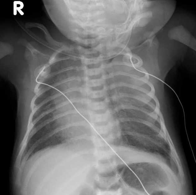
Gastric feeding tube tip in the stomach.
Opacity in the upper zone of the left lung - the differential includes CPAM and a solid mass. A large portion of the left lung is hypolucent.
The film is rotated to the right but nevertheless, the heart and mediastinum appear displaced to the right.
No outstanding finding in the right lung and no pleural effusion.
The heart is normal in size.
The osseous skeleton demonstrated in the film is normal.
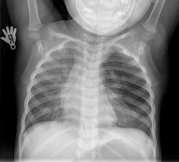
Overinflation of the left upper lobe, displacing the heart and mediastinum to the right.
The right lung is clear.
The heart is normal in size.
Case Discussion
The first chest X-ray on the day of birth was misleading, as it were, as the left upper lobe was radiopaque. In the early neonatal period, the affected lobe contains fetal fluid and tends to be homogenously radiopaque or may show diffuse reticular pattern representing distended lymphatic channels. Subsequent radiograph shows progressive overinflation of the left upper lobe, in keeping with a congenital lobar overinflation.


 Unable to process the form. Check for errors and try again.