
Presentation
Prostate cancer detected on transurethral resection of the prostate (TURP). Prostate-specific antigen (PSA): 4.9 ng/mL.
Patient Data


Findings:
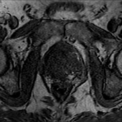
Quality: apex of the prostate cut off in axial T2-weighted and DWI images, does not compromise diagnostic confidence
Prostate size: 71 x 46 x 56 mm (CC x AP x ML) ≈95 mL, PSA density ~0.06 ng/mL2
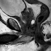
Hemorrhage: diffuse subacute hemorrhage in the apical and midglandular peripheral and transitional zone right>left with inhomogeneously intermediate to low signal on T2-weighted images with interspersed areas of high signal and high signal in T1-weighted images
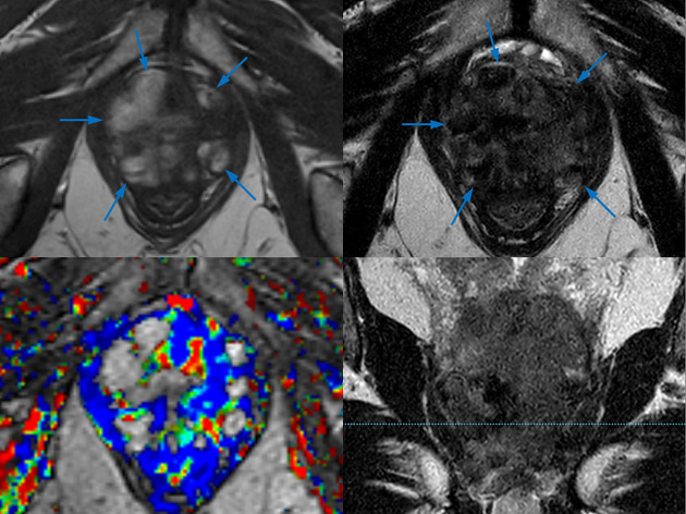
Peripheral zone (PZ): diffusely hypointense bilaterally with broad capsular contact and some residual heterogeneous signal in the right basal and midglandular anterior zones
Transition zone (TZ):
central periurethral defect after transurethral resection of the prostate (TURP)
some BPH nodules in the midglandular anterior transition zone (TZa)
diffuse homogeneous moderate hypointense signal in the midglandular and basal posterior transition zone (TZp) with obscured/indistinct pseudocapsule
Prostate margin: highly irregular with a capsular breach and tumor extension
Neurovascular bundles: asymmetry and invasion of the left neurovascular bundle
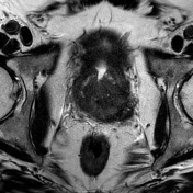
Seminal vesicles: extension of T2w hypointensity and diffusion restriction along the ejaculatory ducts above the base and within the inferior portion of the left seminal vesicle, bleeding in the mid and lateral portions of left seminal vesicle
Lymph nodes: suspicious, enlarged left external iliac lymph node
Pelvic bones: no signs of bony metastasis
Impression:
Histologically proven prostate cancer post transurethral resection of the prostate (TURP), with infiltration of both lobes from basal to apical with extraprostatic extension (EPE) and left seminal vesicle invasion (SVI).
Extensive prostatic hemorrhage predominantly in the apical and posterior midglandular zones.
MRI putative stage: cT3bN1Mx

Key findings:
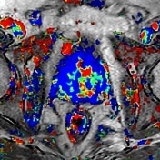
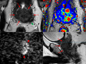
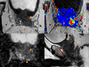
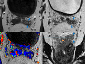
- large prostatic hemorrhage in the apical and midglandular zone of the prostate (blue arrows) - best visible in T1-weighted images, due to the paramagnetic effect of methemoglobin
- extensive tumor growth in the base and midglandular zones of the prostate expressed by the "erased charcoal sign" (red arrows) with correspondent hyperintensity in b1400 images (red arrowheads) with irregular prostatic margins, breaches, and neurovascular bundle asymmetry (green arrowhead)
- tumor invasion of the inferior portion of the left seminal vesicle (orange arrowhead)
- bleeding/hemorrhage in the mid and lateral portion of the left seminal vesicle (blue arrowheads)
Case Discussion
This case shows an extensive bilateral prostate cancer with a large prostatic hemorrhage in a patient, who has had post transurethral resection of the prostate (TURP).
Histology of the transurethral resection of the prostate (TURP) preparation revealed an acinar adenocarcinoma (modified Gleason score 5+4=9, grade 5).
MRI was done for staging and radiation therapy planning.
There is an obvious extraprostatic extension (EPE) and seminal vesicle invasion (SVI). However, there is some confounding, due to large prostatic hemorrhage.
The case also depicts tumor growth and bleeding in the left seminal vesicle.
The patient is scheduled to receive intensity-modulated radiotherapy.


 Unable to process the form. Check for errors and try again.