
Presentation
Hemoptysis.
Patient Data
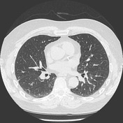

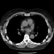
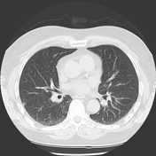
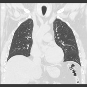
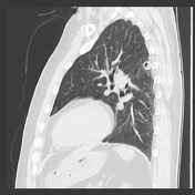
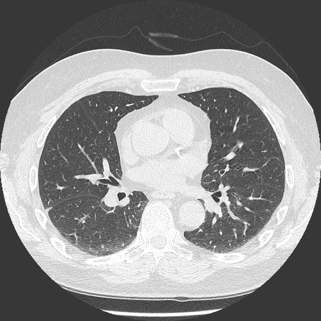
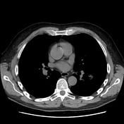
There are bilateral pulmonary nodules measuring up to 2.5 cm in diameter, with one of them, at the superior segment of the left lower lobe, showing cavitation. Biapical pleuro-parenchymal scarring. Lungs and pleural spaces are otherwise clear. Airways are normal. No thoracic lymphadenopathy. Hepatic steatosis partially imaged.
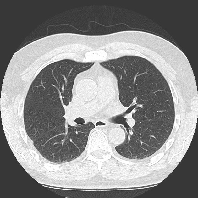
The previously documented pulmonary nodules have improved and mostly resolved. No new nodules.
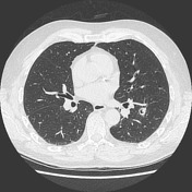

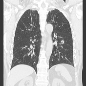
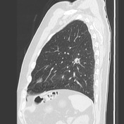

New RUL solid nodule measuring 5 mm and left lower lobe superior segment cavitation nodule measuring 1.5 cm.
Case Discussion
This case illustrates the typical pattern of recurrent/relapsing cavitated pulmonary nodules seen in granulomatosis with polyangiitis. This patient has a confirmed diagnosis and has been in chronic treatment.
Differential for cavitated pulmonary nodules include:
- cavitating pulmonary metastases
- infection (eg TB, cryptococcosis)
- non-infective granulomas:
- granulomatosis with polyangiitis
- rheumatoid nodules


 Unable to process the form. Check for errors and try again.