
Presentation
Presented with cough and breathlessness for 1 week.
Patient Data
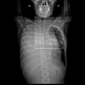


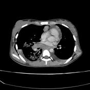
Gross right pleural effusion with complete collapse of right lung. Mediastinal shift to the left side. Left mild pleural effusion. Large soft tissue density mass lesion is seen in the anterior mediastinum extending to superior mediastinum and to the neck on left side. No evidence of calcifications/fat components.

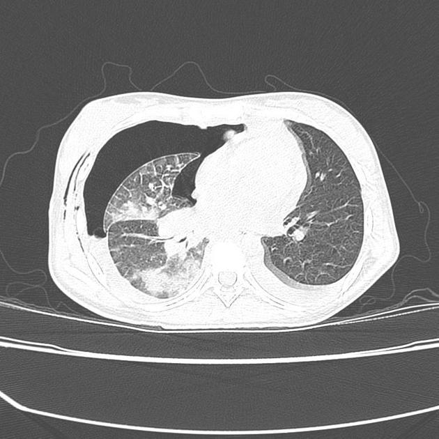
Right hydropneumothorax. ICD tube tip is seen outside the 5th ICS. There is re-expansion of the right lung showing diffuse ground glass opacities and smooth interlobular septal thickening. Left moderate pleural effusion.
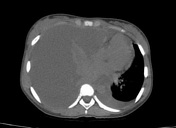
Anterior mediastinal mass shows homogeneous enhancement. The mass lesion is encasing the superior mediastinal vessels without displacement or compression.
Case Discussion
Rapid expansion of the collapsed right lung following drainage of the right pleural effusion resulted in re-expansion pulmonary edema. Imaging features of the anterior mediastinal mass appear to be lymphoma. FNAC revealed highly cellular smear with atypical lymphoid cells.


 Unable to process the form. Check for errors and try again.