
Presentation
A boy with complaints of neck pain worsening at night, associated with gait disturbance due to the right lower limb weakness, starting two months ago. No sensory disturbance.
Patient Data


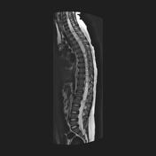
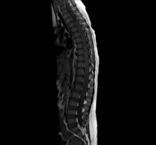
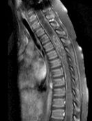
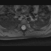
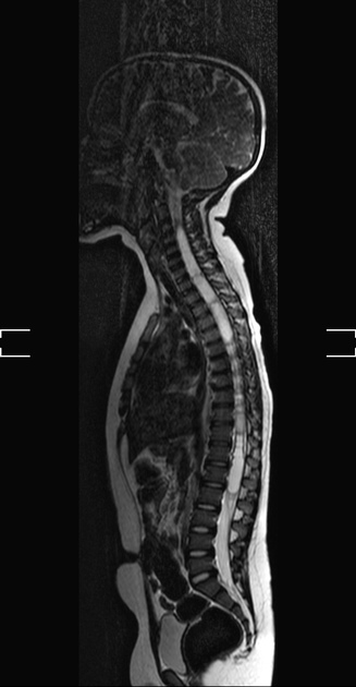
Contrast-enhanced MRI reveals expansile intramedullary lesion between T3 and T6, isointense on T1-WI, hyperintense on T2-WI, with diffuse heterogeneous enhancement. The mass is slightly eccentric to the right side on the axial image, and determines expansion of the thoracic spinal cord, at T3-T6. There is an extensive septated syringohydromyelia, resulting in a thin rim of cervical and thoracic cord, as well as conus medullaris.

Pathology
Macroscopy: In formalin, several irregular, elastic, brownish-gray tissue fragments, measuring the largest 2.5 x 1.6 x 0.6 cm. The cuts are compact.
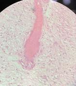
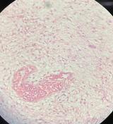
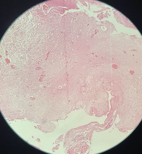
Microscopy: Histology reveals reactive astrocytes with biphasic morphology characterized by a combination of loose glial components consisting of multipolar cells, microcysts, occasional macrocysts, and eosinophilic granular bodies, as well as a more compact piloid tissue with delicate fibrillary areas, cells with long bipolar processes, Rosenthal fibers, and elongated cytologically bland nuclei. There are also hyalinized vessels. The lack of mitotic activity, endothelial proliferation, and necrosis support the histologic diagnosis of a grade I astrocytoma.
Conclusion: Histological pattern compatible with WHO grade I pilocytic astrocytoma.
Case Discussion
Spinal astrocytomas are the most common spinal cord tumor in children 1-4. Spinal pilocytic astrocytomas (WHO I), the most common histologic subtype 1,3,4, is a low-grade glioma with benign biologic behavior, and the possibility of cure, if entirely removed through surgical procedure 1,3,5,6. Magnetic resonance imaging (MRI) is the tool of choice for evaluation of these tumors 1-3,5,6.
This case illustrates the radiological features of a spinal pilocytic astrocytoma, WHO I, with extensive syringohydromyelia. The patient underwent resection of the intramedullary tumor between T3 to T6, with a good evolution in the postoperative period.
Case courtesy
- Gillis Almeida Lago, MD - PGY-3, Radiology Resident, Department of Radiology
- Sterfferson Morais, MD – PGY-3, Radiology Resident, Department of Radiology
- Luciana de Oliveira Borges, MD – Pathologist, Department of Pathology
- Antonio Rodrigues de Aguiar Neto, MD - Radiologist, Department of Radiology
Hospital da Restauração – Recife, PE – Brazil


 Unable to process the form. Check for errors and try again.