
Presentation
Evaluate adenopathy.
Patient Data
Age: 80 years
Gender: Male
From the case:
Diffuse nodal and peritoneal amyloidosis


Download
Info
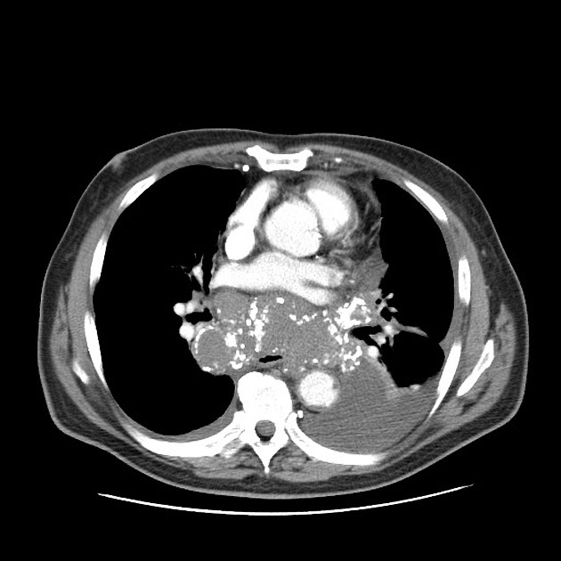
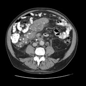
Small right effusion. Medium-large left effusion. Partially calcified pleural plaque. Bulky, partially calcified axillary, mediastinal, retroperioneal, mesenteric, and pelvic adenopathy. Fine nodules/calcifications involving a few areas of the omentum.
Case Discussion
Striking case highlight an uncommon manifestation of systemic amyloidosis (confirmed with axillary LN biopsy), with massive mediastinal, retroperitoneal, and mesenteric adenopathy/amyloidomas. The abdominopelvic disease could be easily mistaken for lymphomatosis or carcinomatosis.


 Unable to process the form. Check for errors and try again.