
Presentation
Abdominal pain and distention.
Patient Data
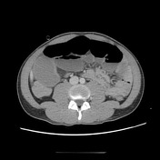


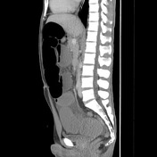

There are wall thickening and mural hyperenhancement involving the terminal ileum for about 14 cm in extension. Focal stricture at the end of this segment leads to upstream small bowel dilatation. Note is also made to antimesenteric pseudosacculation and prominence of the vasa recta within this inflamed segment of the small bowel. Surrounding inflammatory fat stranding but no discrete abscess or fistula. The appendix and part of the cecum appear to have some prominent enhancement, unclear if primary involvement of Crohn's or secondary to the surrounding inflammatory process. NGT in situ.
Small amount of free fluid in the peritoneal cavity, no free gas.
Case Discussion
This case illustrates features of active Crohn disease involving the terminal ileus associated with stricture that leads to the small bowel obstruction presentation - upstream small bowel loops have caliber over 4 cm.
This patient is known to the gastroenterology unit with a known diagnosis of Crohn disease.


 Unable to process the form. Check for errors and try again.