
Presentation
Staging CT. Multiple hemorrhagic cerebral metastases.
Patient Data
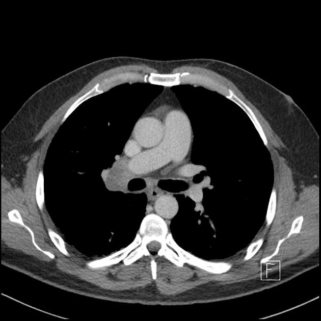
Enlarged right upper paratracheal and precarinal nodal masses with central hypodensity. Multiple other smaller mediastinal lymph nodes noted. Enlarged right hilar lymph node is narrowing the right upper lobe pulmonary artery. The precarinal and hilar nodal masses are compressing on the azygos vein at this confluence with the SVC. Filling defect within the azygos vein represents a thrombus. No left hilar lymphadenopathy.
No central pulmonary arterial filling defects. RUL nodule. No pleural effusion.
No concerning osseous lesions.
IMPRESSION
Mediastinal and right hilar lymphadenopathy with RUL nodule - likely primary bronchogenic malignancy with nodal metastases.
Azygos vein thrombosis.
Case Discussion
Histology of right parietal lesion: Metastatic poorly differentiated carcinoma with morphologic features in keeping with adenocarcinoma. Although TTF-1 is negative, the immune profile would be consistent with metastasis of lung origin.


 Unable to process the form. Check for errors and try again.