
Presentation
Left shoulder swelling.
Patient Data
Age: 6 years
Gender: Female
From the case:
Ewing sarcoma

Download
Info
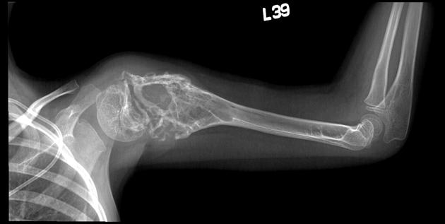
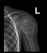
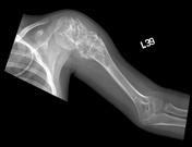
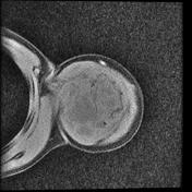
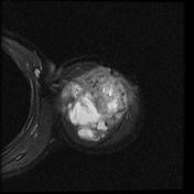
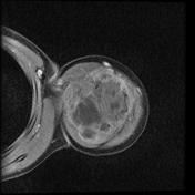
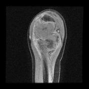
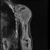
Large expansile mass involving the proximal humeral metaphysis.

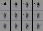
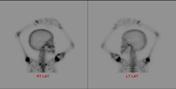
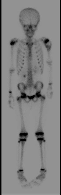
From the case:
Ewing sarcoma


Download
Info
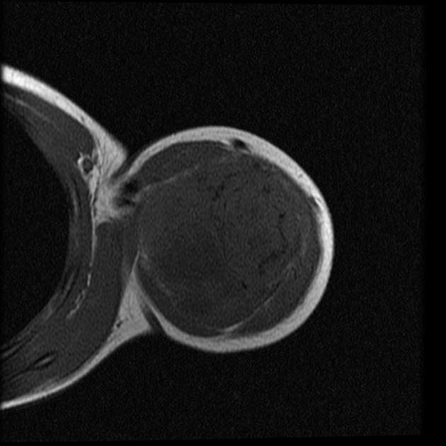
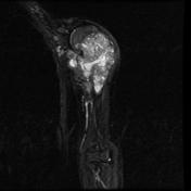
MRI not only confirms the presence of a mass but also demonstrates a large extra-osseous component both laterally beneath deltoid, as well as medially into the axilla. The mass is isointense to muscle on T1, heterogeneously high on T2 and demonstrates extensive contrast enhancement.
Case Discussion
Pathologically proven Ewing sarcoma.


 Unable to process the form. Check for errors and try again.