Presentation
Infant presenting with forceful vomiting.
Patient Data


Ultrasound of the pylorus demonstrates 4mm thick pyloric muscle and increased length of the pyloric canal to 20mm. The thickened muscle forms a prominent hypoechoic target sign on transverse (sagittal with respect to the trunk) imaging . Also note the protrusion of pyloric mucosa in to the antrum.
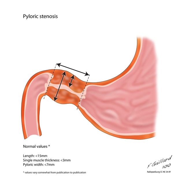
In a normal situation, the pyloric muscle thickness (diameter of a single muscular wall on a transverse image) should normally be less than 3 mm (most accurate) and the length (longitudinal measurement) should not exceed 15 mm.
Case Discussion
In the correct clinical scenario the diagnosis of pyloric stenosis stenosis can be made with confidence on the basis of ultrasound alone.
Case courtesy of Bob Cook, MD. Western Memorial Regional Hospital Corner Brook, Newfoundland.


 Unable to process the form. Check for errors and try again.